©The Author(s) 2017.
World J Gastrointest Surg. Dec 27, 2017; 9(12): 281-287
Published online Dec 27, 2017. doi: 10.4240/wjgs.v9.i12.281
Published online Dec 27, 2017. doi: 10.4240/wjgs.v9.i12.281
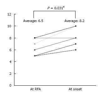
Figure 1 Child-Pugh score significantly increased between “just after radiofrequency ablation” to at the onset of perforation (P = 0.
031). aIndicates values that are statistically significant (P < 0.05). RFA: Radiofrequency ablation.
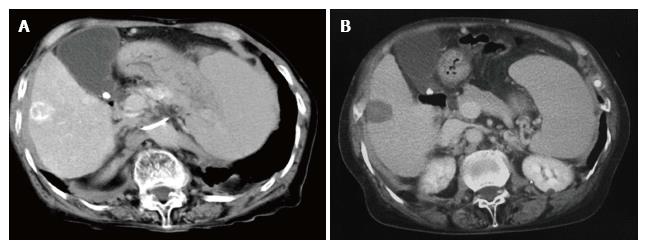
Figure 2 Tumors treated by radiofrequency ablation.
A: Contrast-enhanced computed tomography (CT) shows hepatocellular carcinoma in segment 6 of the liver (Case 4); B: Abdominal CT image at just radiofrequency ablation shows a lesion of ablation (Case 4).
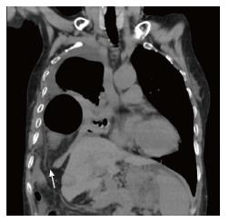
Figure 3 Coronal computed tomography image at onset of diaphragm perforation, showing a right diaphragm hernia.
The right colon is deviated into the thoracic cavity through the diaphragm defect (white arrow) (Case 4).
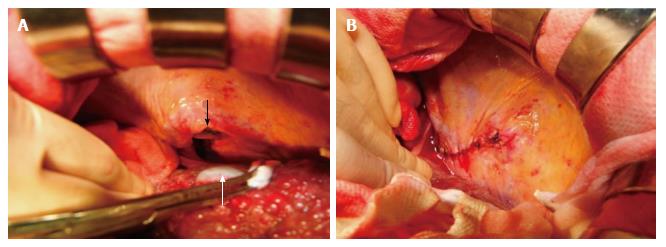
Figure 4 All cases of diaphragm rupture were treated by surgical laparotomy and simple suture of the diaphragm defect.
A: A 5 cm defect of diaphragm is visible (black arrow), with evidence of post-ablation scarring (white arrow) (Case 2); B: The defect was repaired with interrupted sutures (Case 2).
- Citation: Nagasu S, Okuda K, Kuromatsu R, Nomura Y, Torimura T, Akagi Y. Surgically treated diaphragmatic perforation after radiofrequency ablation for hepatocellular carcinoma. World J Gastrointest Surg 2017; 9(12): 281-287
- URL: https://www.wjgnet.com/1948-9366/full/v9/i12/281.htm
- DOI: https://dx.doi.org/10.4240/wjgs.v9.i12.281


