©The Author(s) 2016.
World J Gastrointest Surg. Jun 27, 2016; 8(6): 467-471
Published online Jun 27, 2016. doi: 10.4240/wjgs.v8.i6.467
Published online Jun 27, 2016. doi: 10.4240/wjgs.v8.i6.467
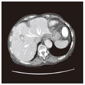
Figure 1 Right hepatic artery branch aneurysm.
Transversal view on contrast enhanced abdominal computed tomography exhibiting a 10 mm × 6 mm aneurysm originating from a branch of the right hepatic artery irrigating segment VIII. The darker halo surrounding the aneurysm reflects secondary hepatic parenchyma lower perfusion due to mass effect. The lithiasic gallbladder is not seen on this image.
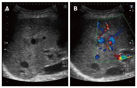
Figure 2 Aneurysm on hepatic Doppler-ultrasound.
Sagittal view of the liver representing the aneurysm at the center of the echographic field (A) and Doppler signals confirming the blood flow inside the aneurysm (B). The branch from the hepatic artery the aneurysm is originating from is not seen on the image.
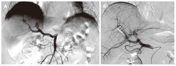
Figure 3 Supra-selective angiography of the hepatic artery branch aneurysm.
A: The left image is before embolization; it shows the hepatic artery splitting and the aneurysm taking source from one of the right branches; B: The right image is after supra-selective embolization of the aneurysm with microcoils. The catheter is still visible. Note that the hepatic artery originates from the superior mesenteric artery. This exhibits a full hepatic artery variant, type IX in Michels’ classification.
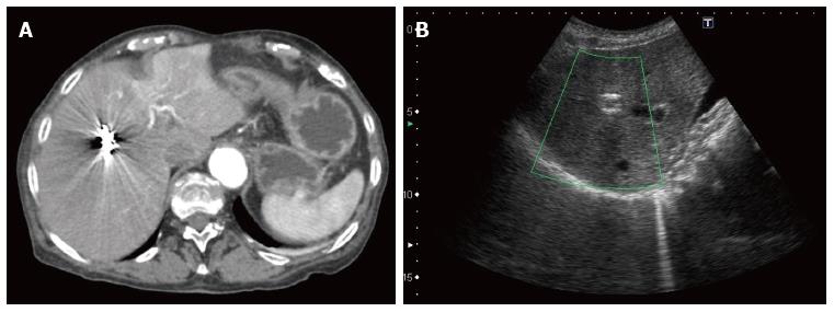
Figure 4 Post embolization imaging.
A: Transversal view on the contrast enhanced abdominal computed tomography performed two days after embolization showing that the coils are in place and the absence of blood extravasation; B: Thirty-days control liver ultrasound showing coils in place in the sagittal plane.
- Citation: Vultaggio F, Morère PH, Constantin C, Christodoulou M, Roulin D. Gastrointestinal bleeding and obstructive jaundice: Think of hepatic artery aneurysm. World J Gastrointest Surg 2016; 8(6): 467-471
- URL: https://www.wjgnet.com/1948-9366/full/v8/i6/467.htm
- DOI: https://dx.doi.org/10.4240/wjgs.v8.i6.467


