©The Author(s) 2016.
World J Gastrointest Surg. Apr 27, 2016; 8(4): 326-334
Published online Apr 27, 2016. doi: 10.4240/wjgs.v8.i4.326
Published online Apr 27, 2016. doi: 10.4240/wjgs.v8.i4.326
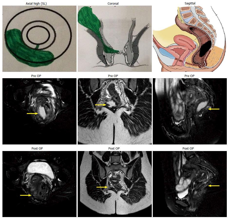
Figure 1 Intersphincteric supralevator abscess and fistula.
A 25-year-old female with supralevator collection from 5 to 9 o’clock. Postoperative magnetic resonance images after 6 wk (bottom row) show complete disease resolution. SL: Supralevator; OP: Operation.
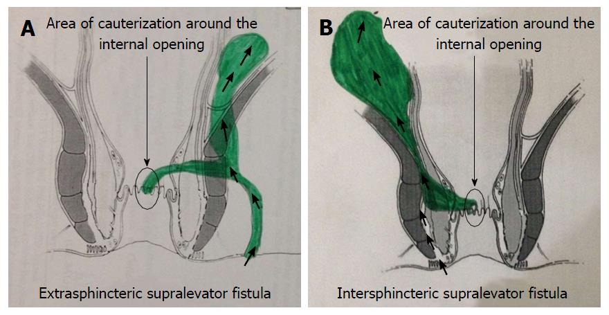
Figure 2 Approach to curette the supralevator fistula.
A: Extrasphincteric; B: Intersphincteric.
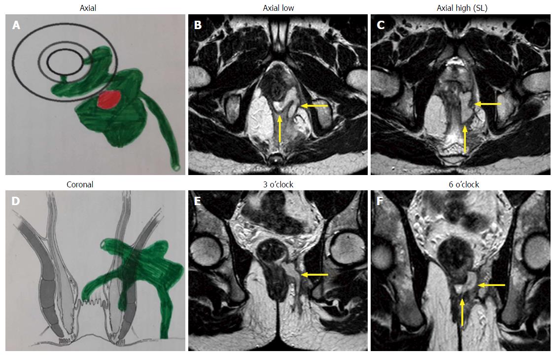
Figure 3 Transsphincteric supralevator fistula.
A 22-year-old male patient with infralevator posterior fistula opening at 6 o’clock and supralevator opening at 3 o’clock. A-C: Axial; D-F: Coronal.
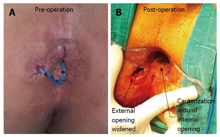
Figure 4 Cauterization around the internal opening and widening of external opening.
Loose draining seton (blue color) can be seen in the preoperative (A) photograph which was inserted during the previous operation by another surgeon 3 mo before the PERFACT procedure was done. A: Preoperatively; B: Postoperatively.
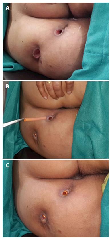
Figure 5 Widened external opening in a patient with multiple tracts (A), removal of tube to clean tracts in the office (B) and reinserted tubes in the tracts after the cleaning process (C).
- Citation: Garg P. PERFACT procedure to treat supralevator fistula-in-ano: A novel single stage sphincter sparing procedure. World J Gastrointest Surg 2016; 8(4): 326-334
- URL: https://www.wjgnet.com/1948-9366/full/v8/i4/326.htm
- DOI: https://dx.doi.org/10.4240/wjgs.v8.i4.326


