Copyright
©2013 Baishideng Publishing Group Co.
World J Gastrointest Surg. Jul 27, 2013; 5(7): 229-232
Published online Jul 27, 2013. doi: 10.4240/wjgs.v5.i7.229
Published online Jul 27, 2013. doi: 10.4240/wjgs.v5.i7.229
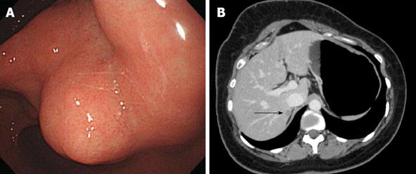
Figure 1 Esophagogastroduodenoscopy.
A: Gastroendoscopy revealed an antral submucosal tumor without ulceration and hemorrhage; B: Computed tomography revealed a hypoattenuating nodule in segment VII of the liver, with peripheral weak enhancement in the portal phase (arrow).
Figure 2 The patient underwent total gastrectomy, regional lymphadenectomy, and partial hepatectomy of segment VII.
A: Macroscopically, gastric tumors were located in the submucosal layer, were round to oval and, relatively well demarcated, and exhibited multiple nodules (n = 8); B: Gastric tumor demonstrated cellular epithelioid gastrointestinal stromal tumors with discohesive pattern of growth (HE, × 40) and mild nuclear atypia (hematoxylin and eosin, × 200) (lower right), exhibiting diffuse cytoplasmic membranous immunoreactivity for CD117 (immunohistochemical stain, × 200) (upper right); C: Hepatic partial resection revealed an oval, well circimscribed, orange yellow, soft, solid tumor, 12 mm × 7 mm (left) and microscopically, synaptophysin-positive, hepatic tumor cells were arranged in nests (so called‘zellballen’) (right), surrounded by S100 protein-positive, sustentacular cells (immunohistochemical stain, × 400).
- Citation: Hong SW, Lee WY, Lee HK. Hepatic paraganglioma and multifocal gastrointestinal stromal tumor in a female: Incomplete Carney triad. World J Gastrointest Surg 2013; 5(7): 229-232
- URL: https://www.wjgnet.com/1948-9366/full/v5/i7/229.htm
- DOI: https://dx.doi.org/10.4240/wjgs.v5.i7.229


