
















©2013 Baishideng Publishing Group Co.
World J Gastrointest Surg. Jun 27, 2013; 5(6): 207-209
Published online Jun 27, 2013. doi: 10.4240/wjgs.v5.i6.207
Published online Jun 27, 2013. doi: 10.4240/wjgs.v5.i6.207
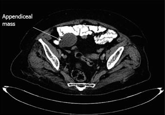
Figure 1 Preoperative contrast enhanced multidetector computed tomography images demonstrated a 4 cm x 3.
5 cm x 3 cm, blind ending, tubular, fluid-filled structure (arrow) that appeared to arise from the cecum, consistent with mucocele of the appendix.
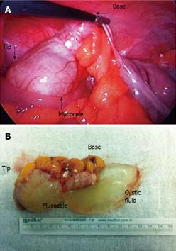
Figure 2 Intra-operative view of smooth cystic tumor of the appendix (A) located at the tip of the appendix (B) after laparoscopic resection (perforated in endobag at the time of extraction).
- Citation: Kaya C, Yazici P, Omeroglu S, Mihmanli M. Laparoscopic appendectomy for appendiceal mucocele in an 83 years old woman. World J Gastrointest Surg 2013; 5(6): 207-209
- URL: https://www.wjgnet.com/1948-9366/full/v5/i6/207.htm
- DOI: https://dx.doi.org/10.4240/wjgs.v5.i6.207