
















©2013 Baishideng Publishing Group Co.
World J Gastrointest Surg. Mar 27, 2013; 5(3): 43-46
Published online Mar 27, 2013. doi: 10.4240/wjgs.v5.i3.43
Published online Mar 27, 2013. doi: 10.4240/wjgs.v5.i3.43
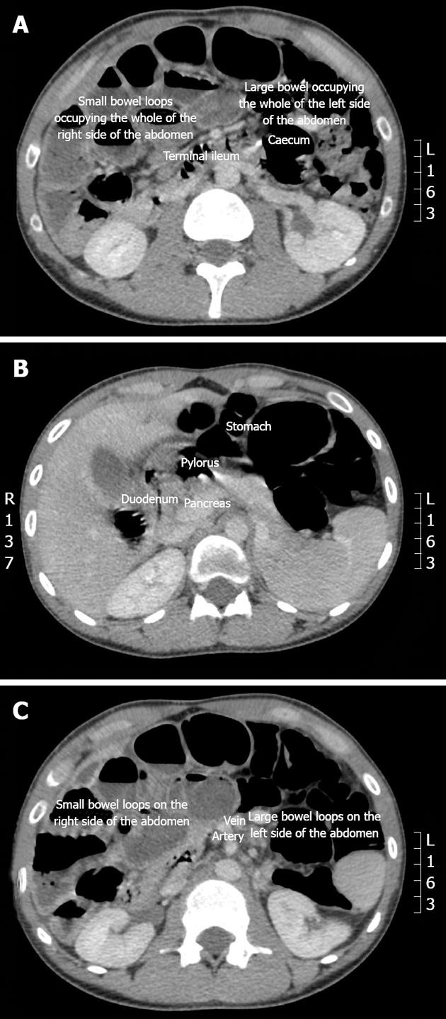
Figure 1 Computerised tomography scan.
A: Abnormal location of the caecum and terminal ileum and most of the small bowel to the right side of the abdomen; B: Non-progression of the duodenum across the spines and aorta; C: Reversal of the relationship between mesenteric artery and vein.
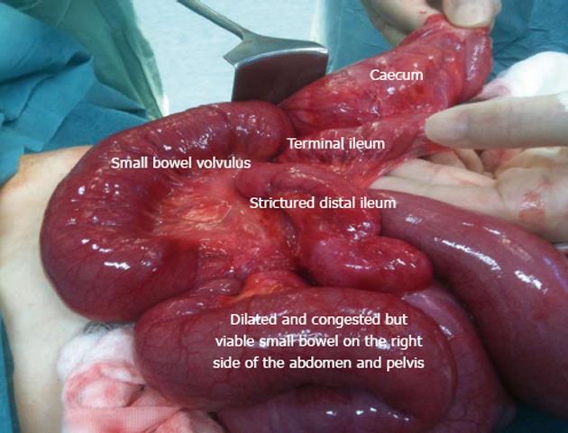
Figure 2 Intraoperative findings showing high riding left upper quadrant caecum, dilated congested but viable small bowel on the right of the abdomen.
Terminal ileum entering the caecum on the right side.
- Citation: Sheikh F, Balarajah V, Ayantunde AA. Recurrent intestinal volvulus in midgut malrotation causing acute bowel obstruction: A case report. World J Gastrointest Surg 2013; 5(3): 43-46
- URL: https://www.wjgnet.com/1948-9366/full/v5/i3/43.htm
- DOI: https://dx.doi.org/10.4240/wjgs.v5.i3.43