©2013 Baishideng Publishing Group Co.
World J Gastrointest Surg. Feb 27, 2013; 5(2): 22-26
Published online Feb 27, 2013. doi: 10.4240/wjgs.v5.i2.22
Published online Feb 27, 2013. doi: 10.4240/wjgs.v5.i2.22
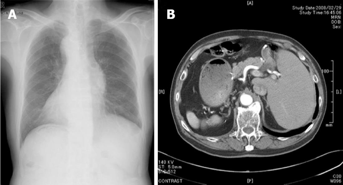
Figure 1 Chest radiography (A) and abdominal computer tomography (B) of the patient.
A: Dextrocardia and a right subphrenic gastric bubble are evident; B: Complete transposition of abdominal viscera is shown, confirming situs inversus totalis.
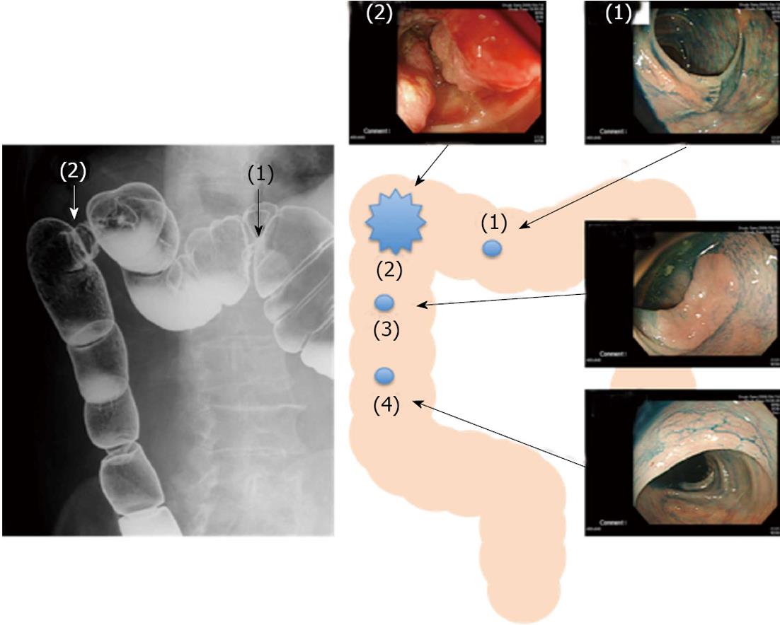
Figure 2 Barium enema and colonoscopy.
Barium enema and colonoscopy showed an ulcerated lesion (2) in the splenic flexure of the transverse colon and a polyp (1) in the transverse colon. Polyps (3, 4) in the descending colon were not detected by barium enema, but were detected by colonoscopy.
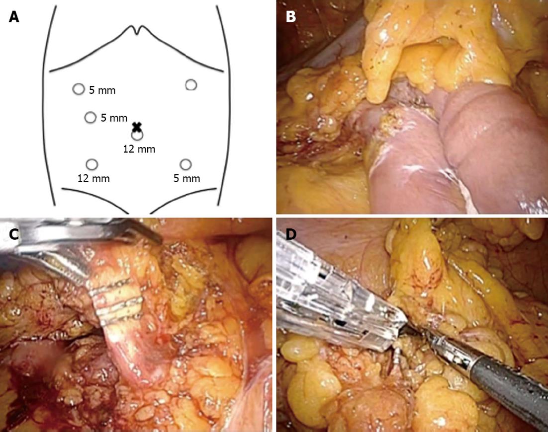
Figure 3 Sites of trocar placement and intraoperative finding.
A: A camera was inserted into the subumbilical area through a 12-mm trocar; B: The reconstruction in the last distal gastrectomy was Billroth II via retrocolic root; C: Radical lymphadenectomy was continued up to the root of the middle colic artery and the branch of the splenic flexure was divided; D: The branch of the right colic artery (the left in an orthotopic patient) was divided.
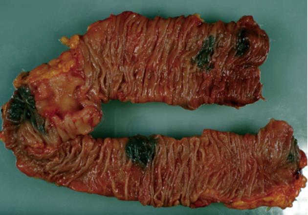
Figure 4 Macroscopic findings.
The main tumor was a 51 mm × 41 mm type 3 lesion in the transverse colon. A 38 mm × 17 mm IIc lesion was in the transverse colon and 17 mm × 13 mm and 10 mm × 10 mm IIa lesions were in the descending colon.
- Citation: Sumi Y, Tomono A, Suzuki S, Kuroda D, Kakeji Y. Laparoscopic hemicolectomy in a patient with situs inversus totalis after open distal gastrectomy. World J Gastrointest Surg 2013; 5(2): 22-26
- URL: https://www.wjgnet.com/1948-9366/full/v5/i2/22.htm
- DOI: https://dx.doi.org/10.4240/wjgs.v5.i2.22


