©2013 Baishideng Publishing Group Co.
World J Gastrointest Surg. Dec 27, 2013; 5(12): 332-336
Published online Dec 27, 2013. doi: 10.4240/wjgs.v5.i12.332
Published online Dec 27, 2013. doi: 10.4240/wjgs.v5.i12.332
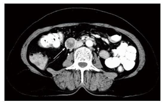
Figure 1 Computed tomography showed a well-demarcated enhancing tumor 4.
0 cm in diameter in the third portion of the duodenum (white arrow).
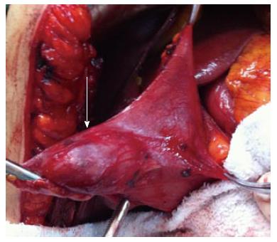
Figure 2 An endophytic gastrointestinal stromal tumor of the third portion of the duodenum (white arrow).
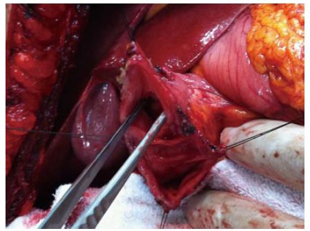
Figure 3 Local limited wedge resection was subsequently performed with clear margins.
Surrounding bowel can be seen to be healthy, allowing for a primary anastomosis.
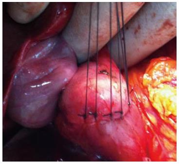
Figure 4 Wedge resection with primary closure.
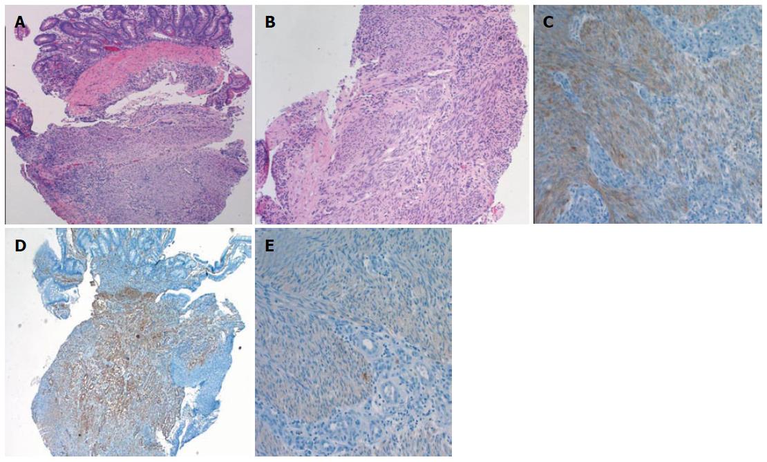
Figure 5 Histology.
A: Submucosal tumor tissue is located (hematoxylin-eosin stain, original magnification, × 5); B: Spindle tumor tissue is composed of cells (hematoxylin-eosin stain, original magnification, × 10); C: Tumor tissue widely seen moderately strong staining of CD117 (CD117, original magnification, × 20); D: Tumor tissue widely seen SMA staining (SMA, original magnification, × 5); E: Tumor tissue, common, poor, S-100 staining is observed (S-100, original magnification, × 20).
- Citation: Acar F, Sahin M, Ugras S, Calisir A. A gastrointestinal stromal tumor of the third portion of the duodenum treated by wedge resection: A case report. World J Gastrointest Surg 2013; 5(12): 332-336
- URL: https://www.wjgnet.com/1948-9366/full/v5/i12/332.htm
- DOI: https://dx.doi.org/10.4240/wjgs.v5.i12.332


