©2013 Baishideng Publishing Group Co.
World J Gastrointest Surg. Oct 27, 2013; 5(10): 264-267
Published online Oct 27, 2013. doi: 10.4240/wjgs.v5.i10.264
Published online Oct 27, 2013. doi: 10.4240/wjgs.v5.i10.264
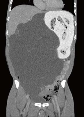
Figure 1 Abdominal computed tomography-scan revealing an enormous cystic process.
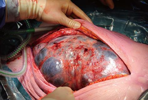
Figure 2 First view after midline incision.
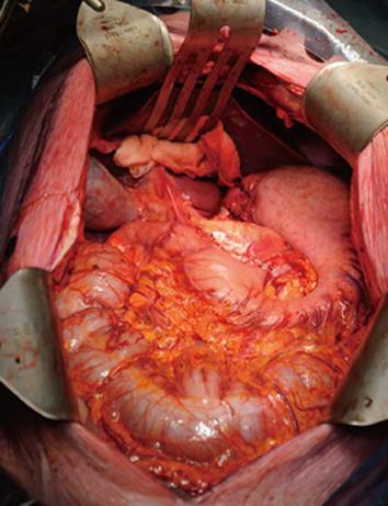
Figure 3 Status after radical resection with partial gastrectomy.
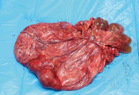
Figure 4 Resected cystic process with near complete fluid drainage.
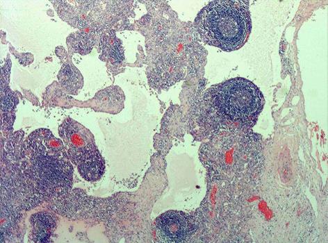
Figure 5 Microscopic view of specimen, depicting various lymphatic spaces (hematoxylin-eosin stain, x 100).
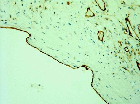
Figure 6 Endothelial lining of cysts (CD31 immunostain, x 400).
- Citation: van Oudheusden TR, Nienhuijs SW, Demeyere TBJ, Luyer MDP, de Hingh IHJT. Giant cystic lymphangioma originating from the lesser curvature of the stomach. World J Gastrointest Surg 2013; 5(10): 264-267
- URL: https://www.wjgnet.com/1948-9366/full/v5/i10/264.htm
- DOI: https://dx.doi.org/10.4240/wjgs.v5.i10.264


