©2012 Baishideng.
World J Gastrointest Surg. Jul 27, 2012; 4(7): 180-184
Published online Jul 27, 2012. doi: 10.4240/wjgs.v4.i7.180
Published online Jul 27, 2012. doi: 10.4240/wjgs.v4.i7.180
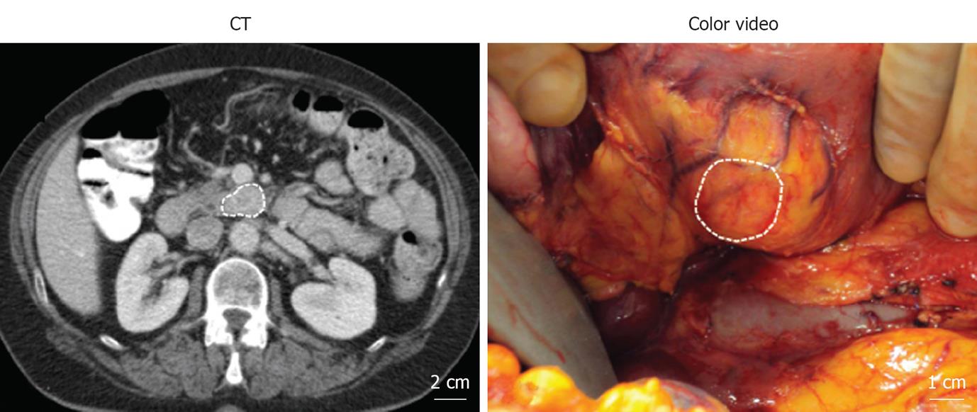
Figure 1 Presurgical and intraoperative visualization of the pancreatic mass.
Figure 2 Mini-FLARE imaging system.
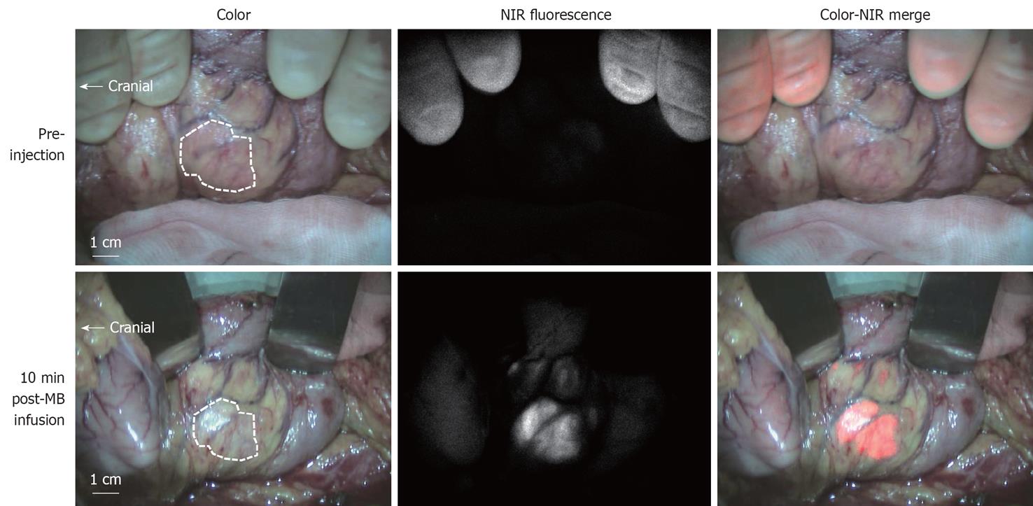
Figure 3 Intraoperative near-infrared fluorescence imaging.
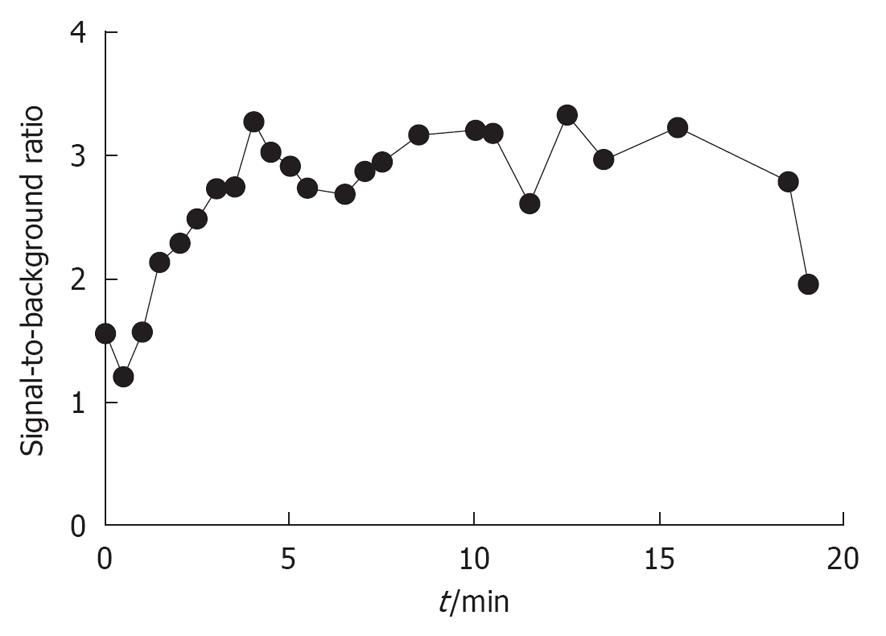
Figure 4 Signal-to-background ratio of the solitary fibrous tumor.
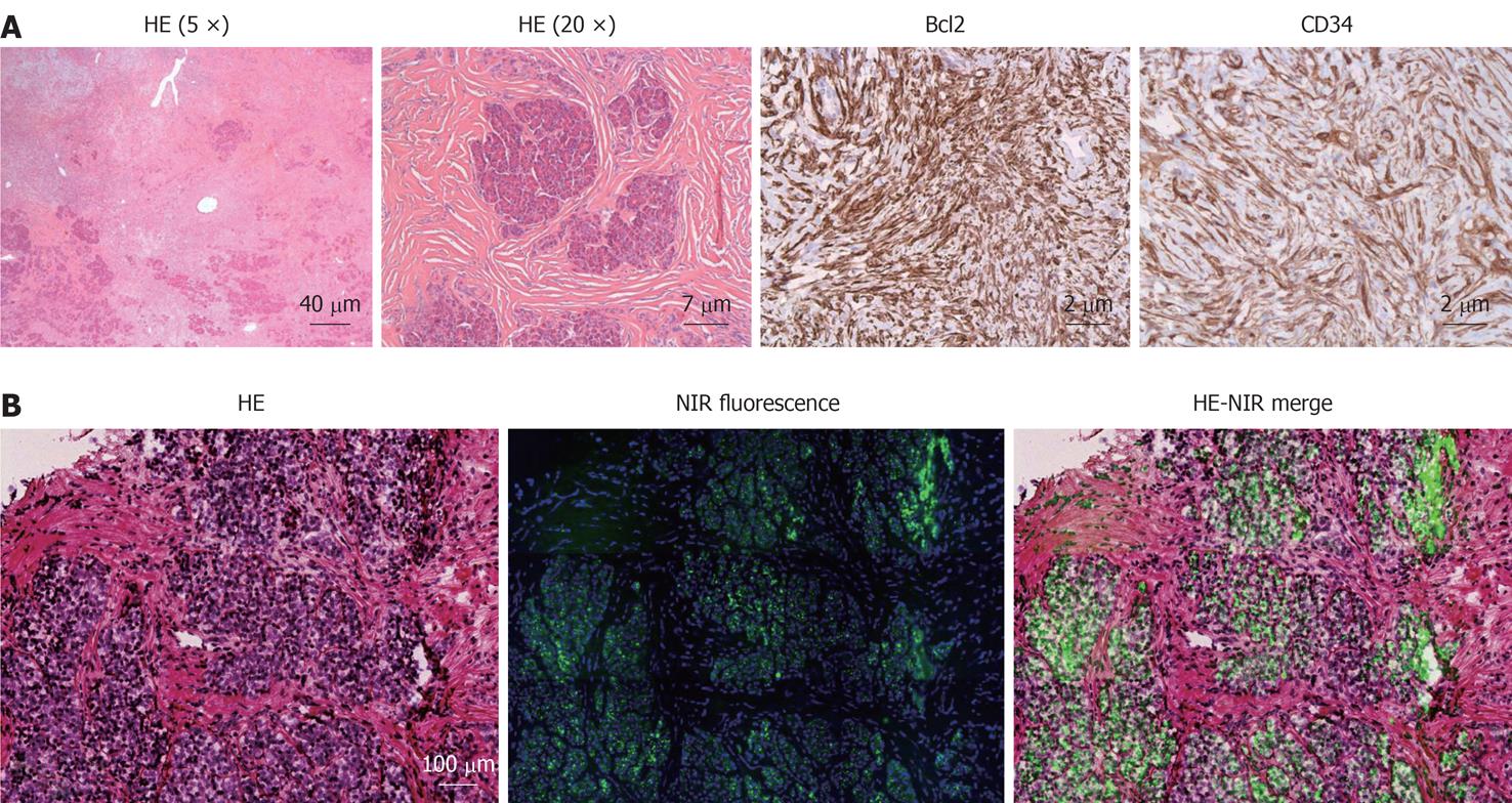
Figure 5 Histopathological evaluation (A) and fluorescence microscopy (B).
- Citation: Vorst JRVD, Vahrmeijer AL, Hutteman M, Bosse T, Smit VT, Velde CJV, Frangioni JV, Bonsing BA. Near-infrared fluorescence imaging of a solitary fibrous tumor of the pancreas using methylene blue. World J Gastrointest Surg 2012; 4(7): 180-184
- URL: https://www.wjgnet.com/1948-9366/full/v4/i7/180.htm
- DOI: https://dx.doi.org/10.4240/wjgs.v4.i7.180


