©2012 Baishideng.
World J Gastrointest Surg. Jul 27, 2012; 4(7): 166-170
Published online Jul 27, 2012. doi: 10.4240/wjgs.v4.i7.166
Published online Jul 27, 2012. doi: 10.4240/wjgs.v4.i7.166
Figure 1 Abdominal computed tomography scan axial (A), sagittal (B) and coronal (C) images of the patient with Grade I pancreatic injury, showing hematoma in the lesser sac and inferomedial displacement of left kidney.
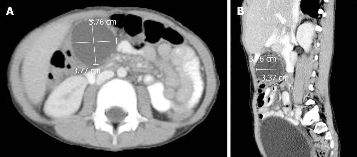
Figure 2 Abdominal computed tomography scan axial (A) and sagittal (B) views showing Grade II pancreatic injury with hematoma measuring 3.
7 cm × 3.7 cm. No other associated injuries were found.
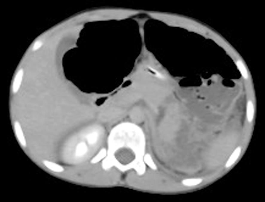
Figure 3 Abdominal computed tomography scan axial view (delayed phase) demonstrating Grade III pancreatic injury with distal transaction of pancreatic tail, dilated loop of transverse colon, and hemoperitoneum.
There is peripancreatic fat stranding as well.
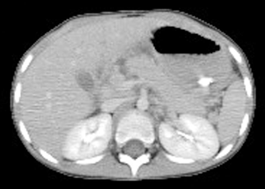
Figure 4 Abdominal computed tomography scan axial view showing Grade V pancreatic injury with shattered head of pancreas.
- Citation: Almaramhy HH, Guraya SY. Computed tomography for pancreatic injuries in pediatric blunt abdominal trauma. World J Gastrointest Surg 2012; 4(7): 166-170
- URL: https://www.wjgnet.com/1948-9366/full/v4/i7/166.htm
- DOI: https://dx.doi.org/10.4240/wjgs.v4.i7.166


