©2012 Baishideng.
World J Gastrointest Surg. May 27, 2012; 4(5): 126-130
Published online May 27, 2012. doi: 10.4240/wjgs.v4.i5.126
Published online May 27, 2012. doi: 10.4240/wjgs.v4.i5.126
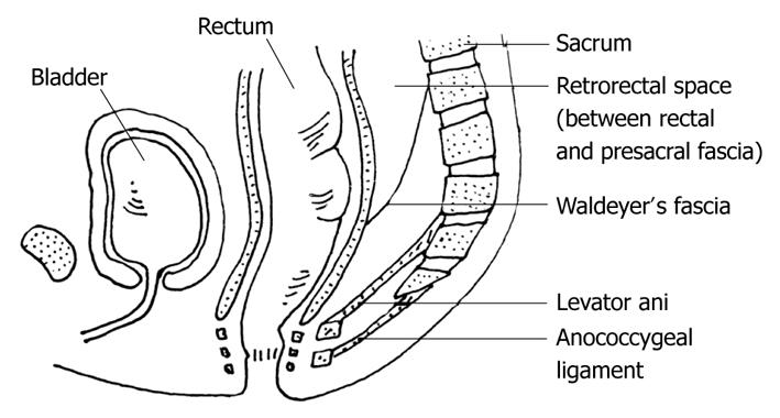
Figure 1 Boundaries of the retrorectal space.
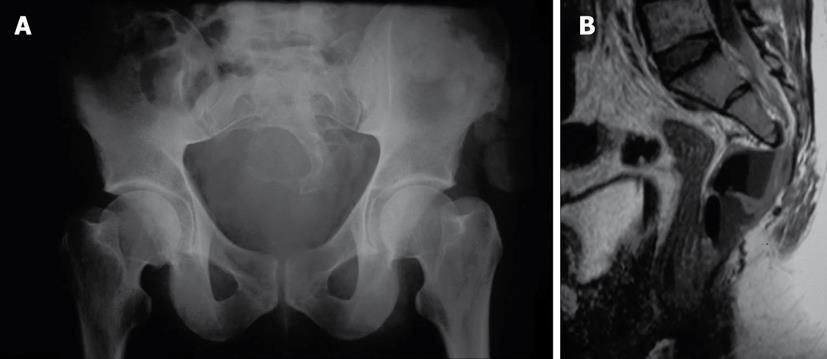
Figure 2 Simple X-ray of the pelvis (A) shows the sacral bony defect (“scimitar sign”) and magnetic resonance imaging (B) established the diagnosis of presacral meningocele, and the presence of air inside suggested the a neuroenteric fistula.
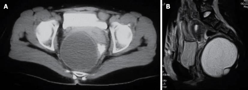
Figure 3 Cystic hamartoma in a 44-year-old woman, computed tomography (A) and magnetic resonance imaging (B).
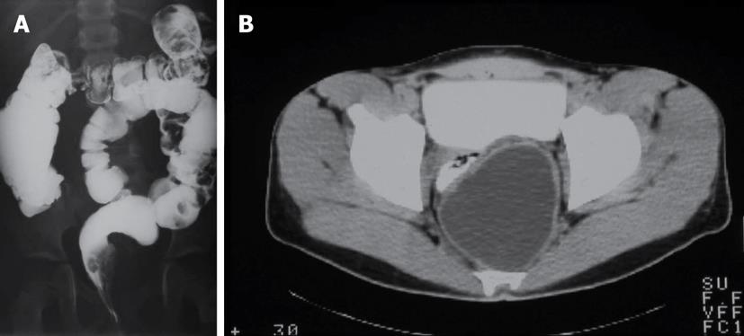
Figure 4 Double-contrast barium enema (A) showing a mass effect in the left wall of the rectum caused by a presacral cystic teratoma (computed tomography, B).
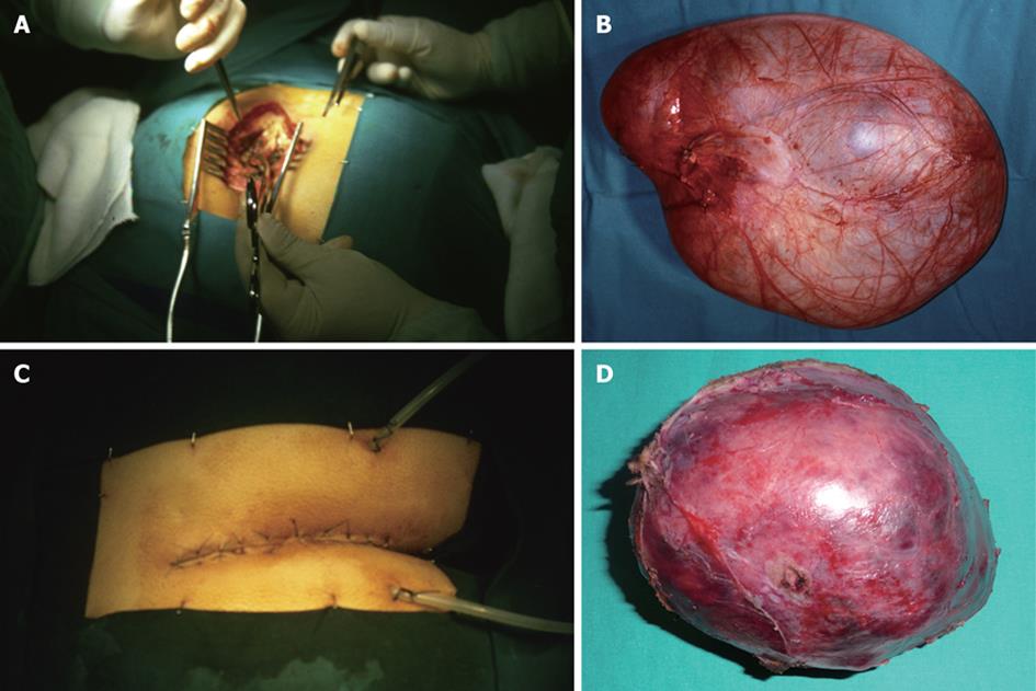
Figure 5 Surgical field (B and D) and specimens (filled with saline solution after complete removal to restore the original volume) (B and D) of patients 2 (A and B) and 3 (C and D).
- Citation: Aranda-Narváez JM, González-Sánchez AJ, Montiel-Casado C, Sánchez-Pérez B, Jiménez-Mazure C, Valle-Carbajo M, Santoyo-Santoyo J. Posterior approach (Kraske procedure) for surgical treatment of presacral tumors. World J Gastrointest Surg 2012; 4(5): 126-130
- URL: https://www.wjgnet.com/1948-9366/full/v4/i5/126.htm
- DOI: https://dx.doi.org/10.4240/wjgs.v4.i5.126


