©2010 Baishideng.
World J Gastrointest Surg. Feb 27, 2010; 2(2): 47-50
Published online Feb 27, 2010. doi: 10.4240/wjgs.v2.i2.47
Published online Feb 27, 2010. doi: 10.4240/wjgs.v2.i2.47
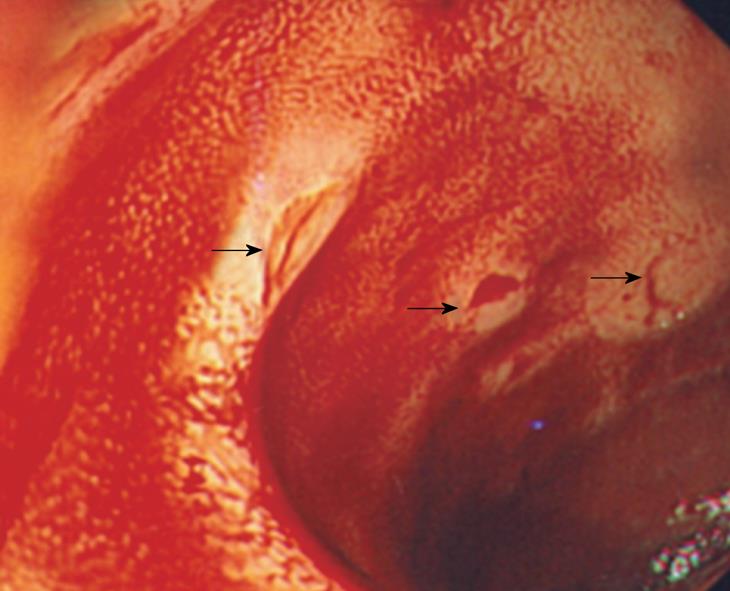
Figure 1 Colonoscopic findings.
Multiple ulcerations with bleeding in terminal ileum (arrows).
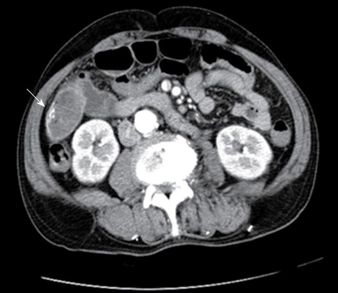
Figure 2 Abdominal CT findings.
Extravasation of contrast dye in small bowel (arrow).
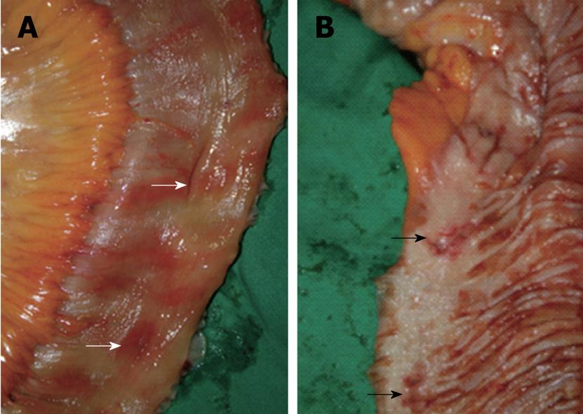
Figure 3 Operative findings.
A: Multiple erythematous lesions in the serosal surface of small bowel (white arrows) are found; B: Multiple ulcerations in the luminal surface of small bowel (black arrows) are noted.
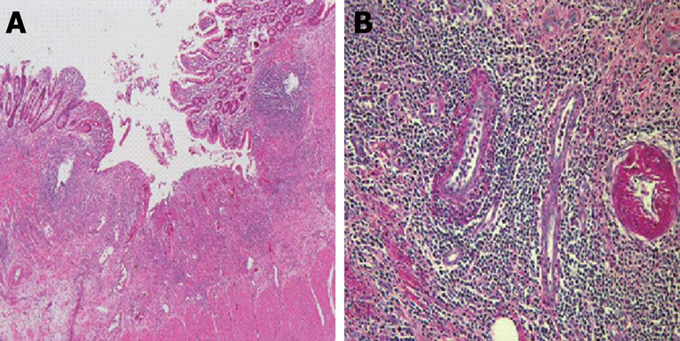
Figure 4 Microscopic Features.
A: Multifocal flask-shaped ulcers are noted (HE × 40); B: Vasculitis is noted with dense infiltration of lymphocytes within the vessel walls in submucosal layer (PAS × 200).
- Citation: Bae KB, Youn WH, Lee YJ, Jung SJ, Hong KH. Massive small bowel bleeding caused by scrub typhus in Korea. World J Gastrointest Surg 2010; 2(2): 47-50
- URL: https://www.wjgnet.com/1948-9366/full/v2/i2/47.htm
- DOI: https://dx.doi.org/10.4240/wjgs.v2.i2.47


