©2010 Baishideng.
World J Gastrointest Surg. Jan 27, 2010; 2(1): 26-29
Published online Jan 27, 2010. doi: 10.4240/wjgs.v2.i1.26
Published online Jan 27, 2010. doi: 10.4240/wjgs.v2.i1.26
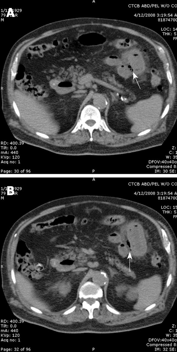
Figure 1 CT scan of a patient with perforated jejunal diverticulitis.
A: The scan demonstrates free air and inflammatory changes surrounding the site of perforation in a patient presenting with left sided focal peritonitis. The arrows point to extra-luminal free air; B: Another transverse image showing free air and inflammatory changes around the site of perforation.
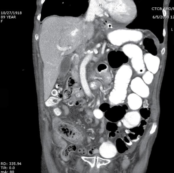
Figure 2 Coronal section of a CT scan demonstrating partial small bowel obstruction (SBO) in a patient with enterolith ileus.
Note the dilated loops of bowel and fecalization just proximal to the obstruction in the right lower quadrant.
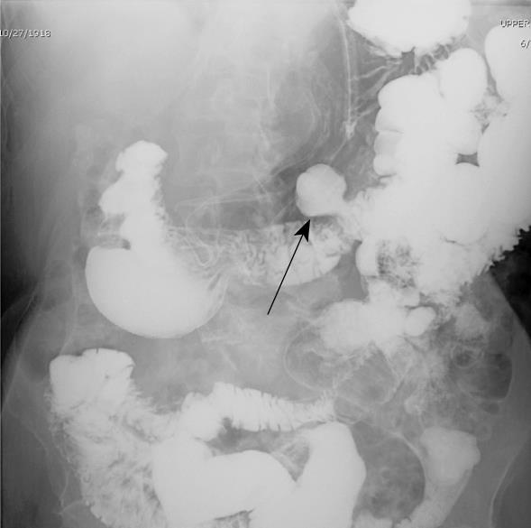
Figure 3 Upper GI with small bowel follow-through demonstrating multiple diverticula in the small bowel as evidenced by the diverticulum (arrow) indicated above.
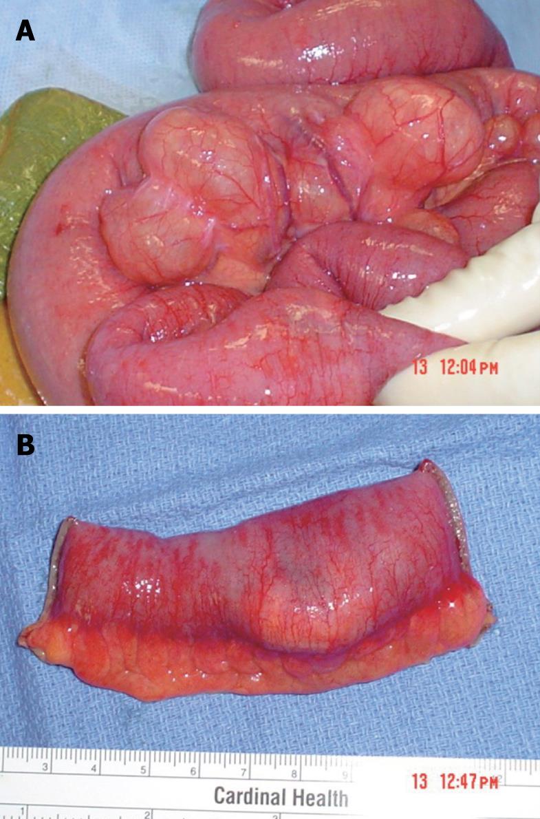
Figure 4 Gross picture of jejunal diverticula disease.
A: Multiple jejunal diverticula are shown; B: Obstructed jejunum from impacted stone with significant hyperemia.
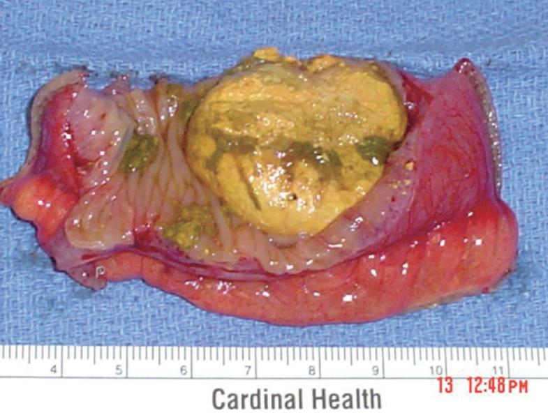
Figure 5 A 3 cm enterolith is noted in the intestine leading to obstruction.
- Citation: Chugay P, Choi J, Dong XD. Jejunal diverticular disease complicated by enteroliths: Report of two different presentations. World J Gastrointest Surg 2010; 2(1): 26-29
- URL: https://www.wjgnet.com/1948-9366/full/v2/i1/26.htm
- DOI: https://dx.doi.org/10.4240/wjgs.v2.i1.26


