
















Copyright: ©Author(s) 2026.
World J Gastrointest Surg. Sep 27, 2026; 18(9): 123459
Published online Sep 27, 2026. doi: 10.4240/wjgs.123459
Published online Sep 27, 2026. doi: 10.4240/wjgs.123459
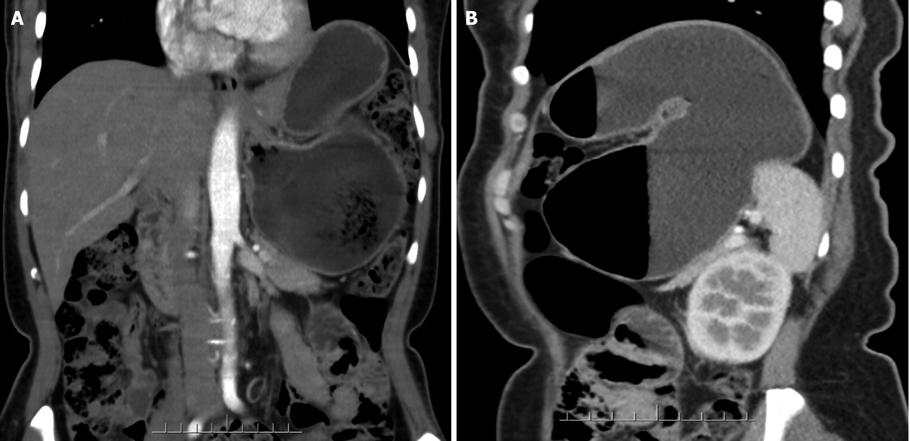
Figure 1 Contrast-enhanced abdominal computed tomography.
A: The coronal view, it shows elevation of the left side of the diaphragm, upward displa
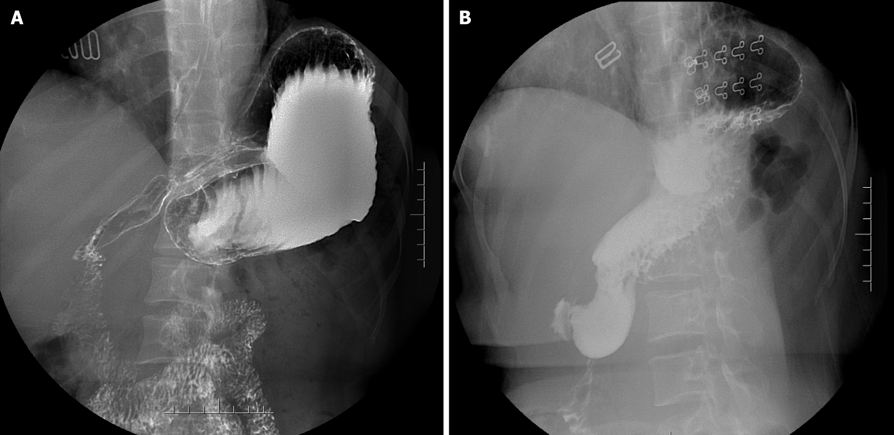
Figure 2 Upper gastrointestinal barium meal before and after surgery.
A: Preoperative: Widening of the upper mediastinal shadow; elevation of the left diaphragm; the stomach is predominantly arched; the fundus is displaced downwards; the body and antrum of the stomach are first displaced upwards and then rotate anti-clockwise to the right; B: Postoperative: Elevation of the left diaphragm; the fundus is displaced more upwards than before; the antrum of the stomach has returned to its normal anatomical position.
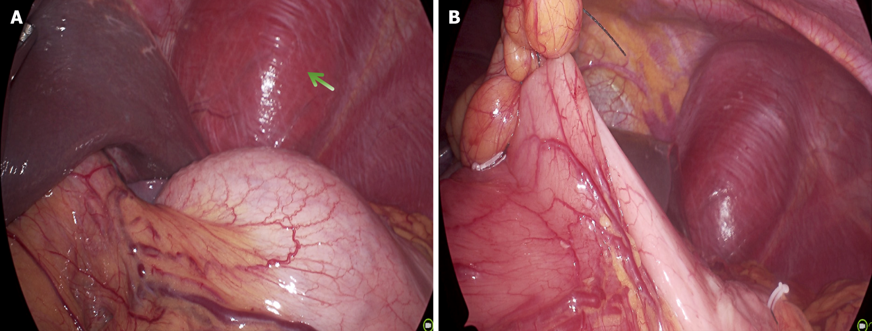
Figure 3 Intraoperative exploration.
A: During the operation, elevation of the left diaphragm was observed; the body of the stomach first twisted from bottom to top, then rotated anti-clockwise towards the lower right. The green arrow indicates the elevation of the left diaphragm; B: During the operation, the greater curvature of the stomach was sutured to the transverse colon, and the anterior wall of the pyloric antrum, near the pylorus, was sutured to the falciform ligament of the liver.
- Citation: Wang S, Yang XF, Wang W. Laparoscopic reduction of gastric volvulus: A case report and review of literature. World J Gastrointest Surg 2026; 18(9): 123459
- URL: https://www.wjgnet.com/1948-9366/full/v18/i9/123459.htm
- DOI: https://dx.doi.org/10.4240/wjgs.123459