
















Copyright: ©Author(s) 2026.
World J Gastrointest Surg. Sep 27, 2026; 18(9): 122838
Published online Sep 27, 2026. doi: 10.4240/wjgs.122838
Published online Sep 27, 2026. doi: 10.4240/wjgs.122838
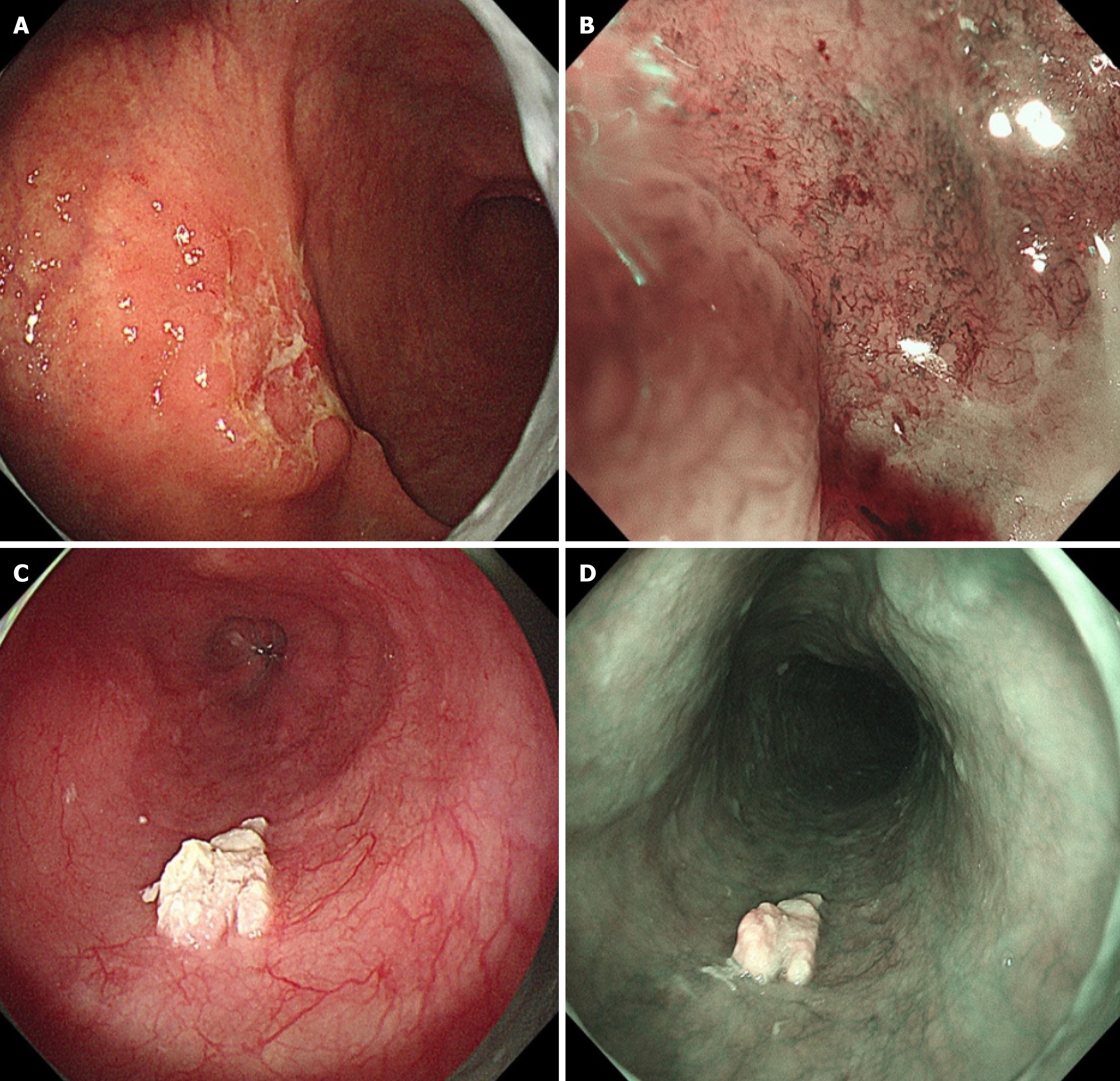
Figure 1 Endoscopic manifestations of gastric and esophageal lesions.
A: White-light endoscopy revealed a Borrmann type II lesion on the greater curvature of the lower stomach body, featuring an ulcer on the surface covered by a moss-like debris; B: Magnifying endoscopy with narrow-band imaging revealed a positive demarcation line, loss of mucosal structure, and an irregular microvascular pattern; C and D: Under white-light and narrow-band imaging, verrucous white patches with clear boundaries are visible; the biopsy specimen from these patches was soft.
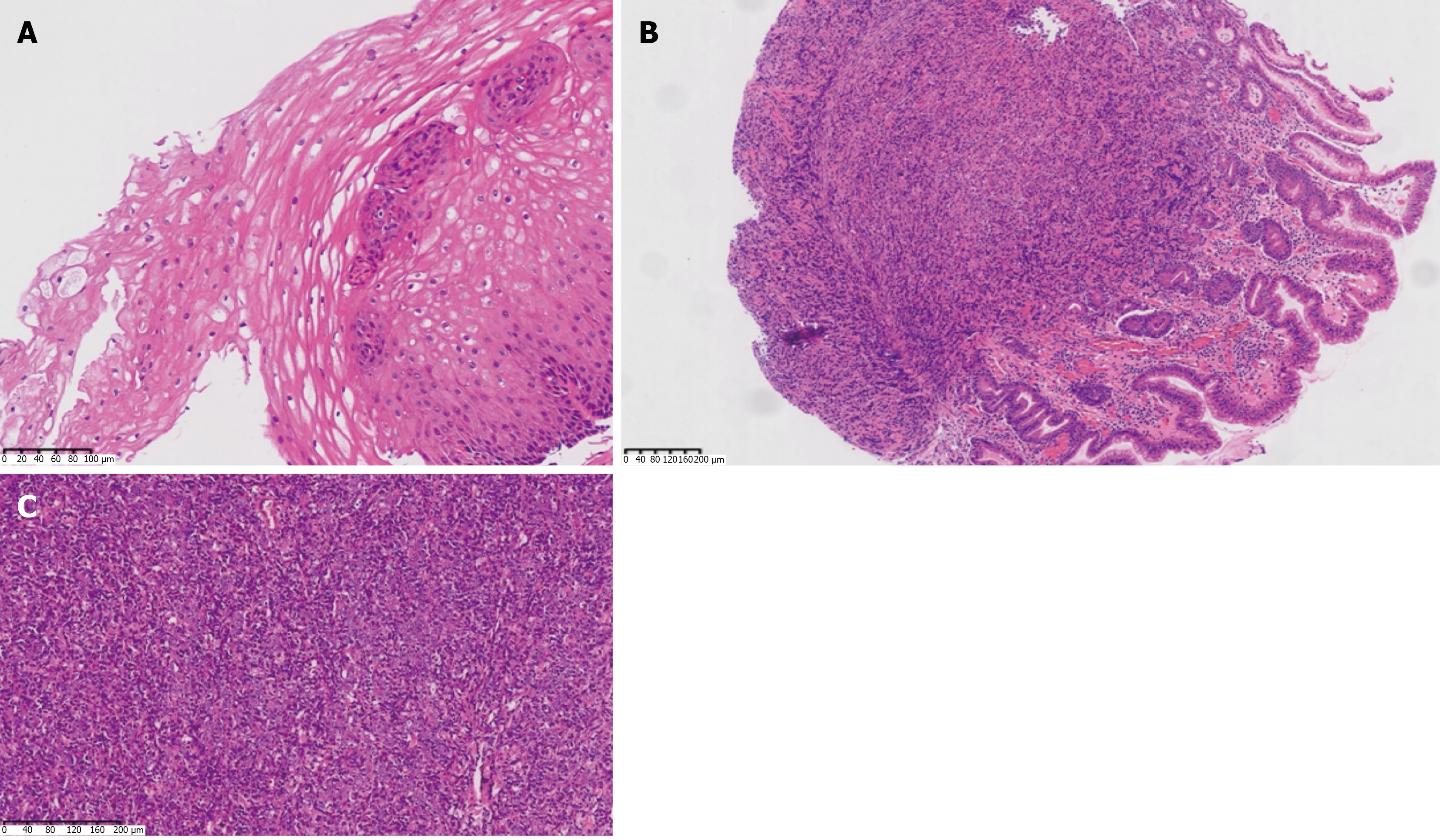
Figure 2 Hematoxylin and eosin-stained histopathological features.
A: The initial esophageal biopsy pathology showing papillary hyperplasia of the squamous epithelium with hyperkeratosis and parakeratosis 31 cm from the incisors; B: The gastric biopsy pathology [4 × hematoxylin and eosin (HE)] showing intramucosal poorly differentiated adenocarcinoma at the greater curvature of the lower gastric body; C: Pathological examination of a radical gastrectomy specimen (10 × HE) showing poorly differentiated adenocarcinoma at the greater curvature of the lower gastric body.
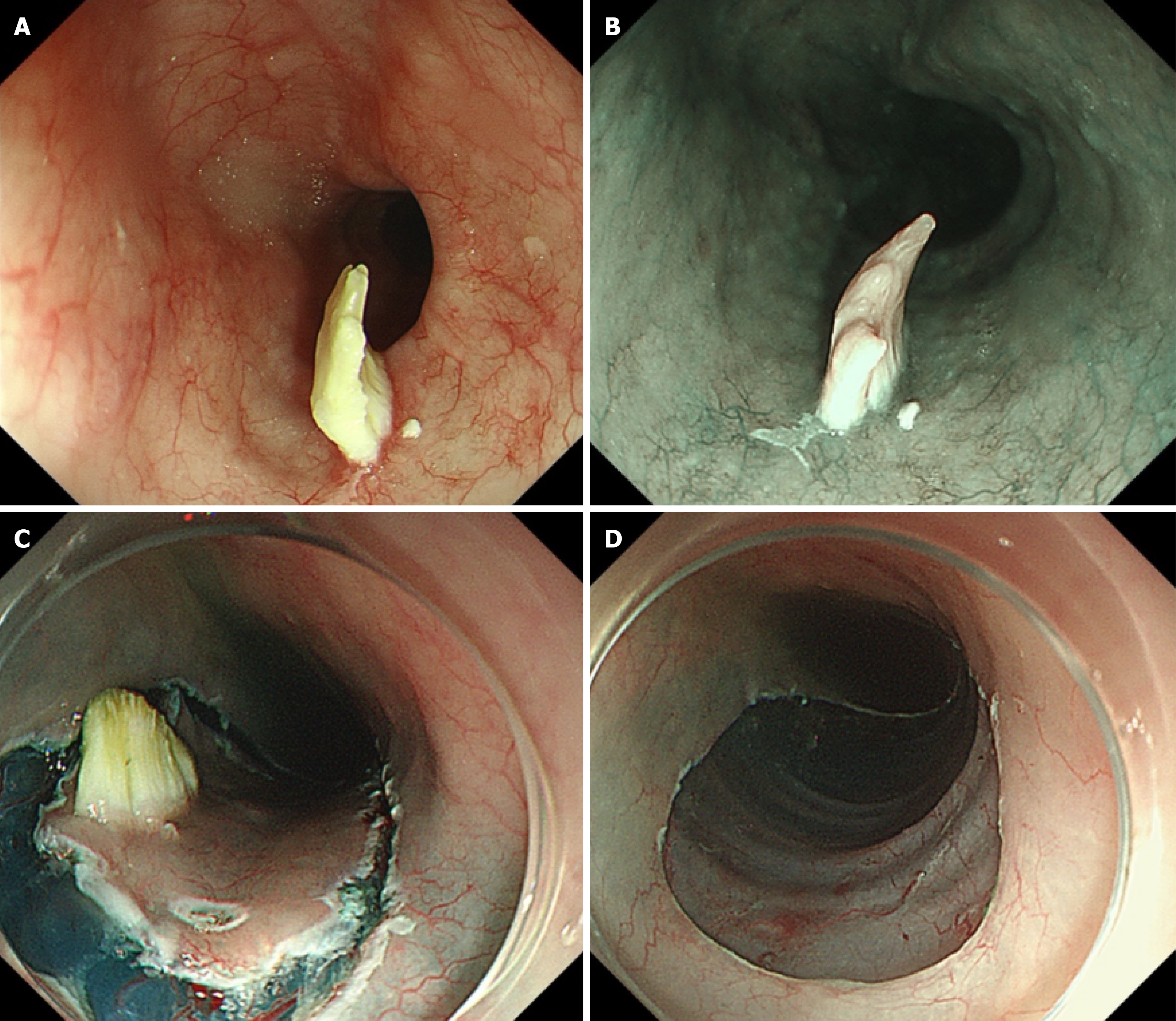
Figure 3 Endoscopic manifestations of the progressive esophageal lesion and the endoscopic submucosal dissection procedure.
A: The lesion appears more protruded than before, showing a whitish and slightly yellowish appearance under white light; B: Narrow-band imaging revealed irregular blood vessels, while the surface structure remained indiscernible; C and D: Endoscopic submucosal dissection was performed to completely excise the lesion.
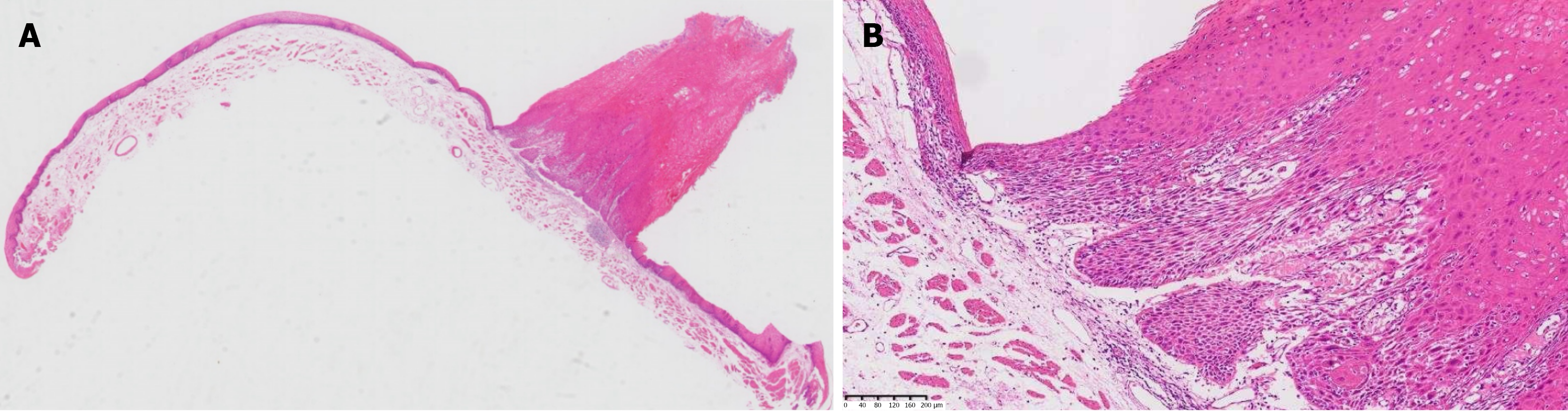
Figure 4 Histopathological features of the esophageal endoscopic submucosal dissection specimen (hematoxylin and eosin staining).
A: Esophageal endoscopic submucosal dissection (ESD) specimen [4 × hematoxylin and eosin (HE)] showing an esophageal mass displaying carcinoma in situ (verrucous or papillary) with a stone pillar-like appearance 31 cm from the incisors; B: Esophageal ESD specimen (10 × HE) showing epithelial cell dysplasia, with no infiltration of the basal cells by the carcinoma in situ.
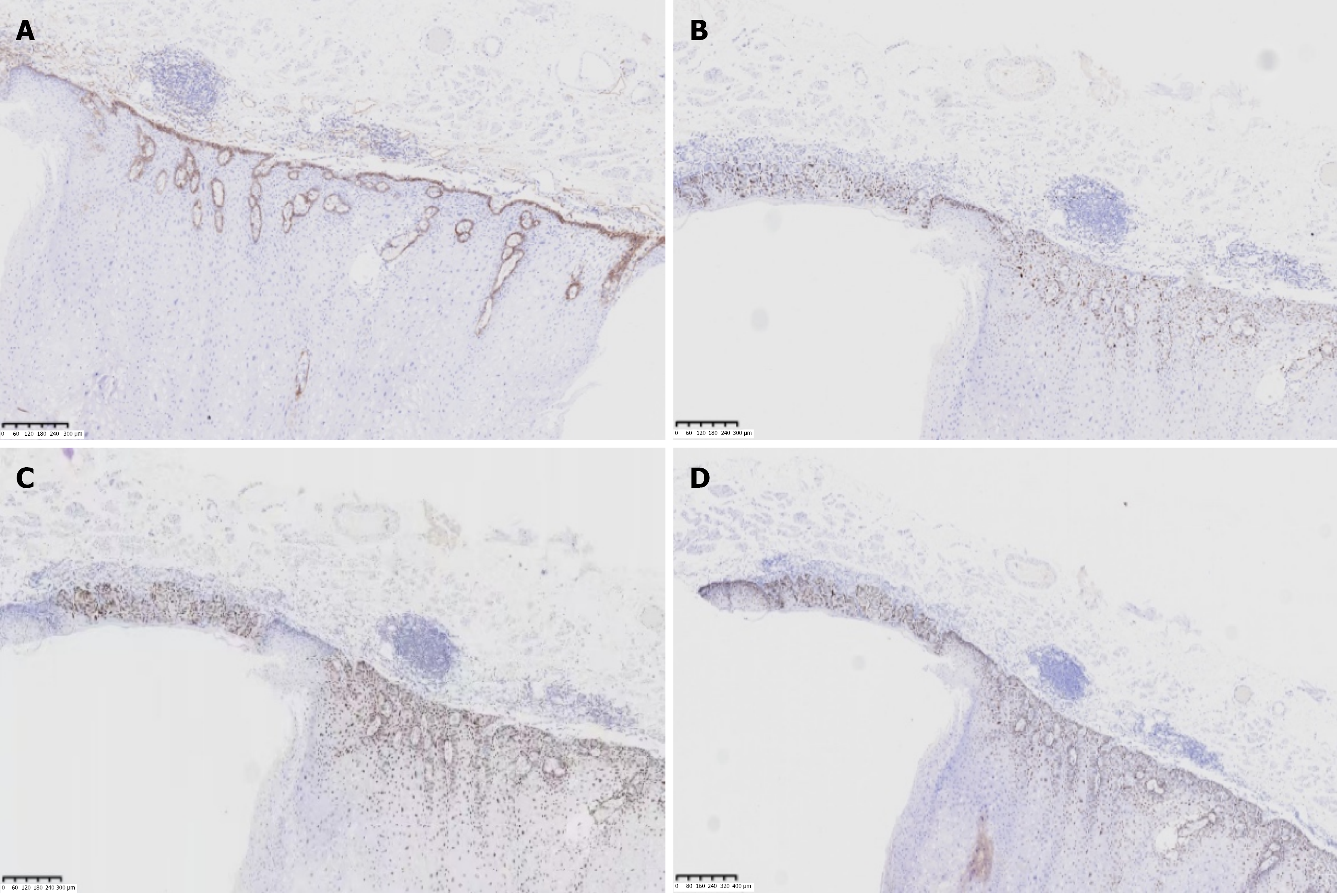
Figure 5 Immunohistochemical staining features of the esophageal endoscopic submucosal dissection specimen.
A: D240 staining shows continuous linear positivity along the membranes of basal cells (clone number: D2–40, positivity was defined as brownish-yellow staining of the basal cell membrane); B: Ki67 staining shows a marked increase in positive cells in the basal layer, with an abnormal distribution pattern [clone number: 176B3C4, abnormal Ki-67 distribution in the squamous epithelium was defined by the presence of any of the following: (1) A higher number of positive cells in the basal layer (≥ 5/HPF); (2) No or significantly reduced expression of cells in the parabasal layer ( < 50%), or an equal or higher number of positive cells in the basal layer compared to the parabasal layer; and (3) The presence of Ki-67 expression in the epithelial superficial layer (> 2 cells/HPF)]; C: P53 staining shows diffuse strong nuclear expression (clone number: 882F5H1, positivity was defined as brownish-yellow staining of the squamous epithelial cell nucleus); D: P63 staining shows positive cytosolic nuclei in tumour cells (clone number: 281B6A9, positivity was defined as brownish-yellow staining of the squamous epithelial cell nucleus).
- Citation: Zhang X, Zhou JY, Wu ZW, Zhou SX, Zheng JF, Zheng WY, Pan WS, Wu HG. Coexistence of esophageal verrucous carcinoma in situ and gastric intramucosal poorly differentiated adenocarcinoma: A case report. World J Gastrointest Surg 2026; 18(9): 122838
- URL: https://www.wjgnet.com/1948-9366/full/v18/i9/122838.htm
- DOI: https://dx.doi.org/10.4240/wjgs.122838