
















Copyright: ©Author(s) 2026.
World J Gastrointest Surg. Sep 27, 2026; 18(9): 121000
Published online Sep 27, 2026. doi: 10.4240/wjgs.121000
Published online Sep 27, 2026. doi: 10.4240/wjgs.121000
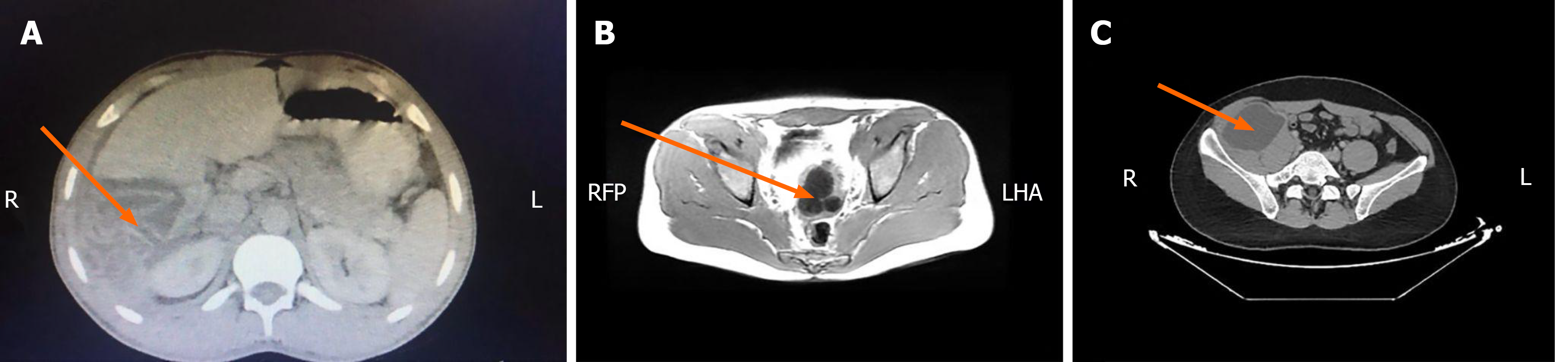
Figure 1 Axial contrast-enhanced computed tomography images demonstrating rare extrahepatic and extrapulmonary hydatid cyst localizations.
A: A well-defined hypodense cystic lesion located within the right adrenal gland (arrow), without evidence of adjacent organ invasion; B: A round, homogeneous cystic mass in the presacral/retrovesical pelvic compartment (arrow), producing mild mass effect on surrounding structures; C: A well-circumscribed unilocular cystic lesion adjacent to the right iliopsoas muscle (arrow), without calcification or daughter cyst formation.
- Citation: Pülat H, Öztaşan D, Akyüz C, Özer BV, Yüksel C. Surgical management and recurrence analysis in primary extrahepatic-extrapulmonary hydatid cysts: A single-center experience of 20 cases. World J Gastrointest Surg 2026; 18(9): 121000
- URL: https://www.wjgnet.com/1948-9366/full/v18/i9/121000.htm
- DOI: https://dx.doi.org/10.4240/wjgs.121000