
















Copyright: ©Author(s) 2026.
World J Gastrointest Surg. Sep 27, 2026; 18(9): 120658
Published online Sep 27, 2026. doi: 10.4240/wjgs.120658
Published online Sep 27, 2026. doi: 10.4240/wjgs.120658
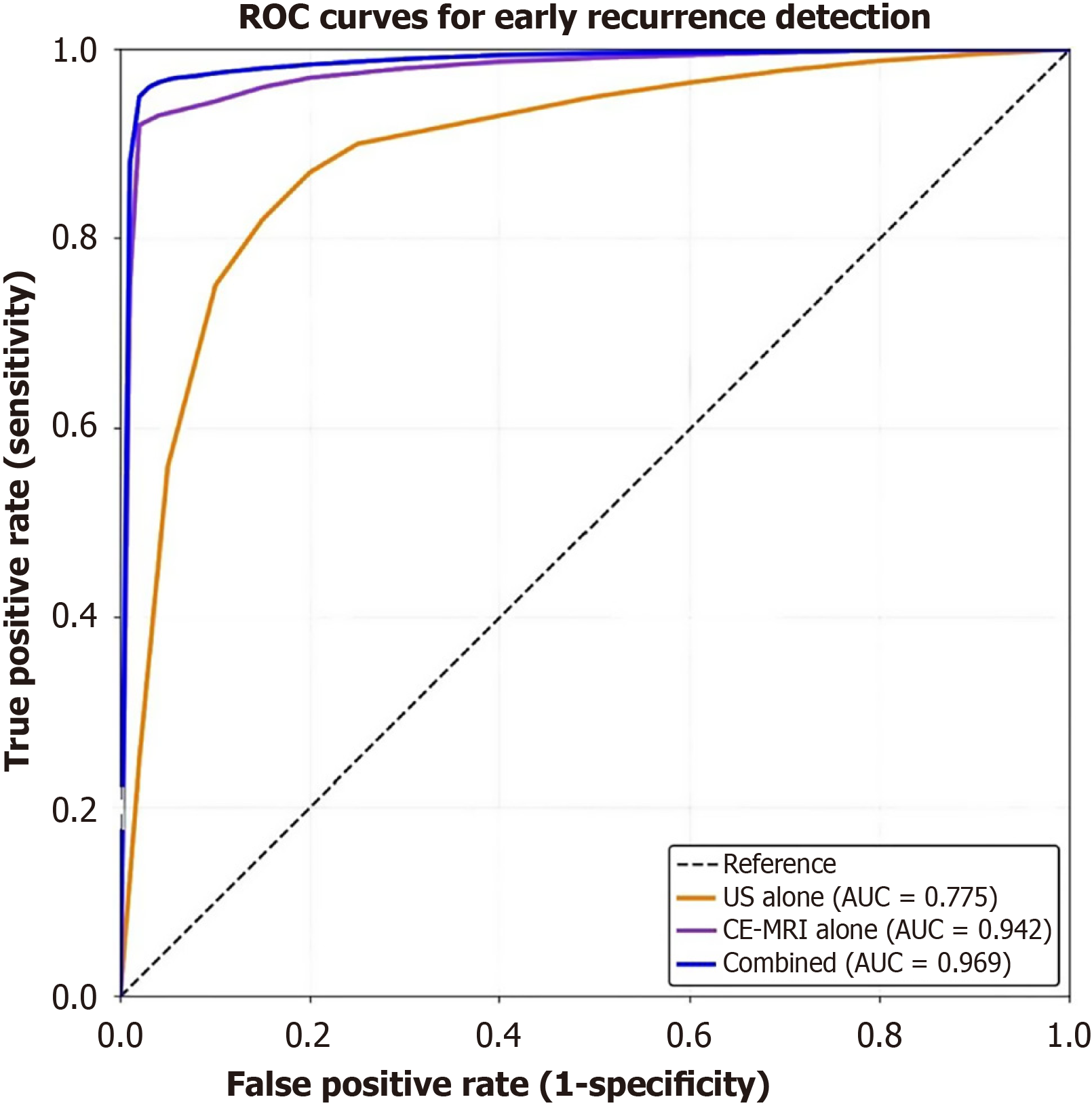
Figure 1 Receiver operating characteristic curves comparing diagnostic performance.
Receiver operating characteristic (ROC) curves demonstrating the diagnostic performance of conventional ultrasound (US), contrast-enhanced magnetic resonance imaging (CE-MRI), and the combined approach for early recurrence detection after hepatocellular carcinoma resection. The combined approach [blue line, area under the curve (AUC) = 0.969] showed significantly superior performance compared with CE-MRI alone (purple line, AUC = 0.942, P = 0.035) and US alone (orange line, AUC = 0.775, P < 0.001). CE-MRI also demonstrated significantly better performance than US (P < 0.001).
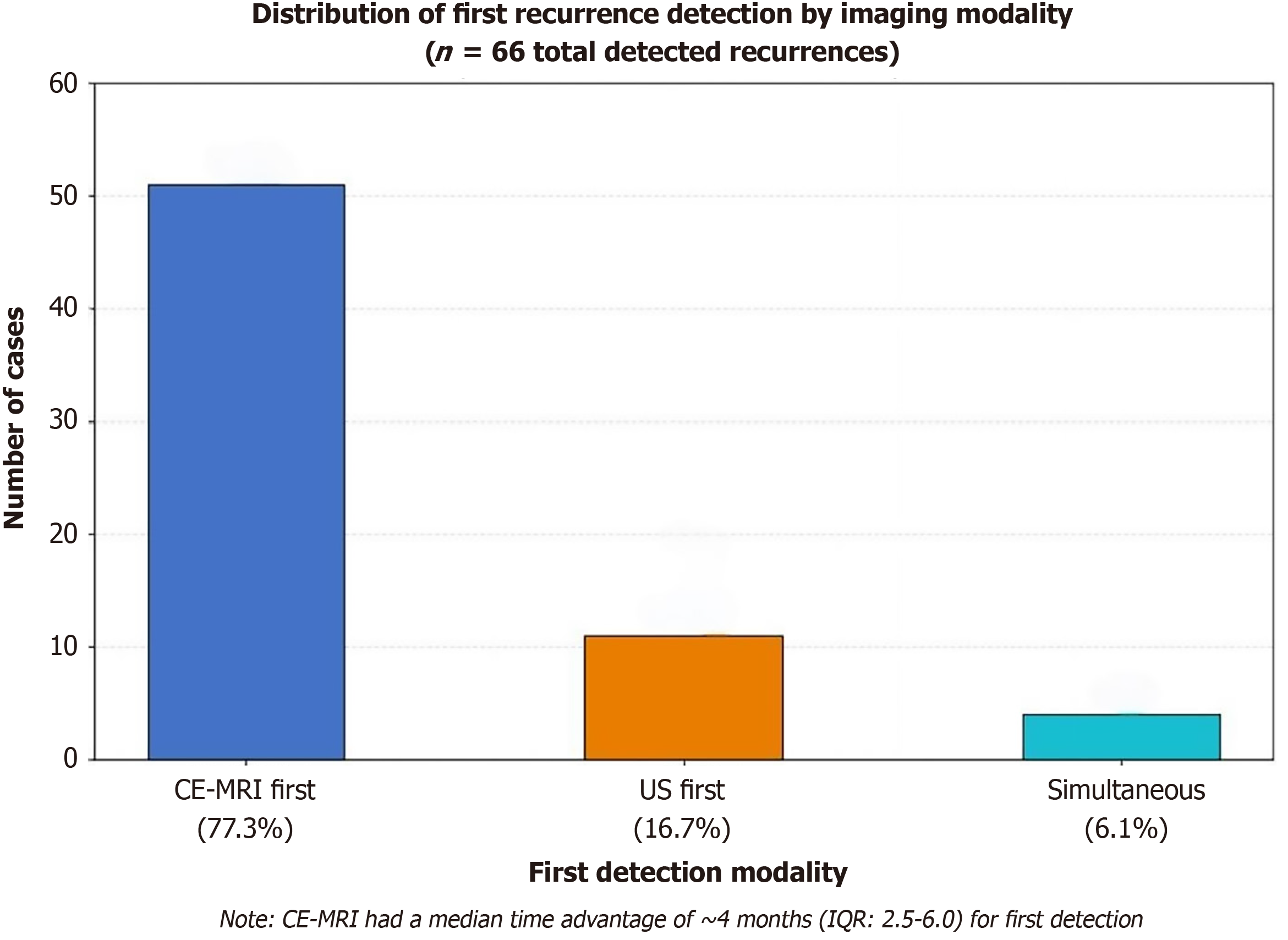
Figure 2 Time distribution of first recurrence detection by imaging modality.
Bar chart showing which imaging modality first detected recurrence among the 66 cases detected by the combined approach. Contrast-enhanced magnetic resonance imaging (CE-MRI) was the first modality to detect recurrence in 51 cases (77.3%, blue bar), ultrasound (US) was first in 11 cases (16.7%, orange bar), and both modalities detected recurrence simultaneously in 4 cases (6.1%, light blue bar). The median time advantage of CE-MRI for first detection was 3.2 months (range, 1.5-6.0 months) compared with subsequent detection by US. IQR: Interquartile range.
- Citation: Deng T, Li XR. Complementary diagnostic value of conventional ultrasound and contrast-enhanced magnetic resonance imaging for post-hepatectomy hepatocellular carcinoma surveillance. World J Gastrointest Surg 2026; 18(9): 120658
- URL: https://www.wjgnet.com/1948-9366/full/v18/i9/120658.htm
- DOI: https://dx.doi.org/10.4240/wjgs.120658