
















Copyright: ©Author(s) 2026.
World J Gastrointest Surg. Sep 27, 2026; 18(9): 120497
Published online Sep 27, 2026. doi: 10.4240/wjgs.120497
Published online Sep 27, 2026. doi: 10.4240/wjgs.120497
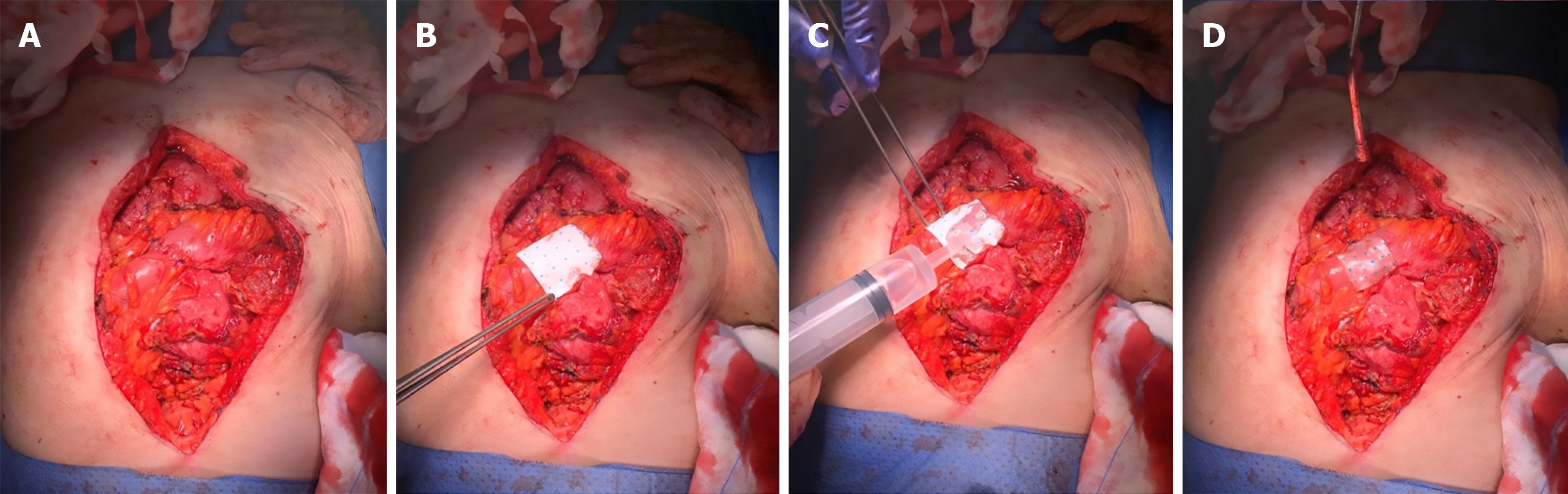
Figure 1 Intraoperative application of Hemopatch® during manual bowel anastomosis.
A: Completed hand-sewn bowel anastomosis; B: Placement of Hemopatch® over the anastomotic line to reinforce the suture; C: Activation of the sealing layer using sodium bicarbonate solution; D: Final adherence of the hydrated Hemopatch® to the anastomosis.
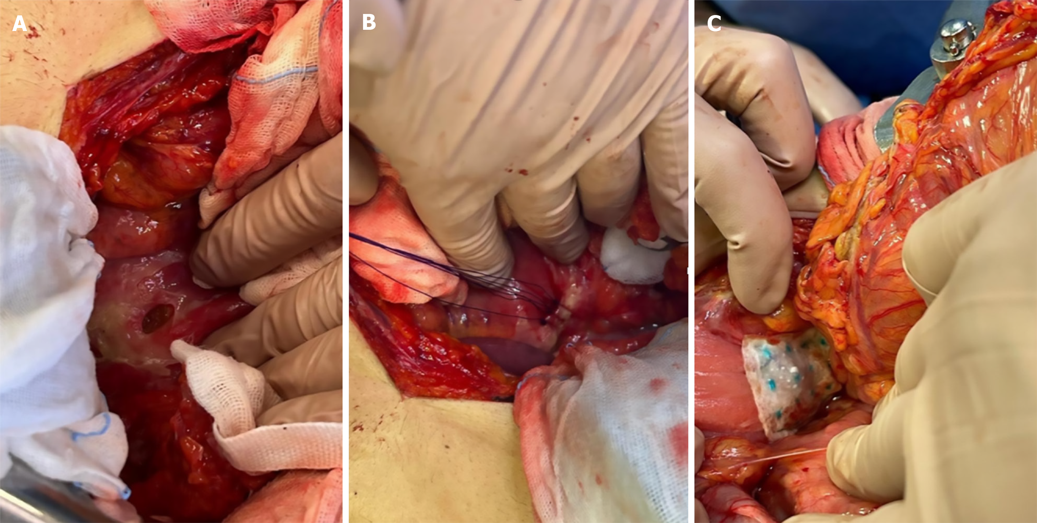
Figure 2 Surgical management of perforated gastric ulcer with reinforcement using Hemopatch®.
A: Intraoperative view of a perforated gastric ulcer; B: Primary closure of the perforation with interrupted sutures; C: Application of Hemopatch® over the suture line to reinforce the repair and provide additional sealing.
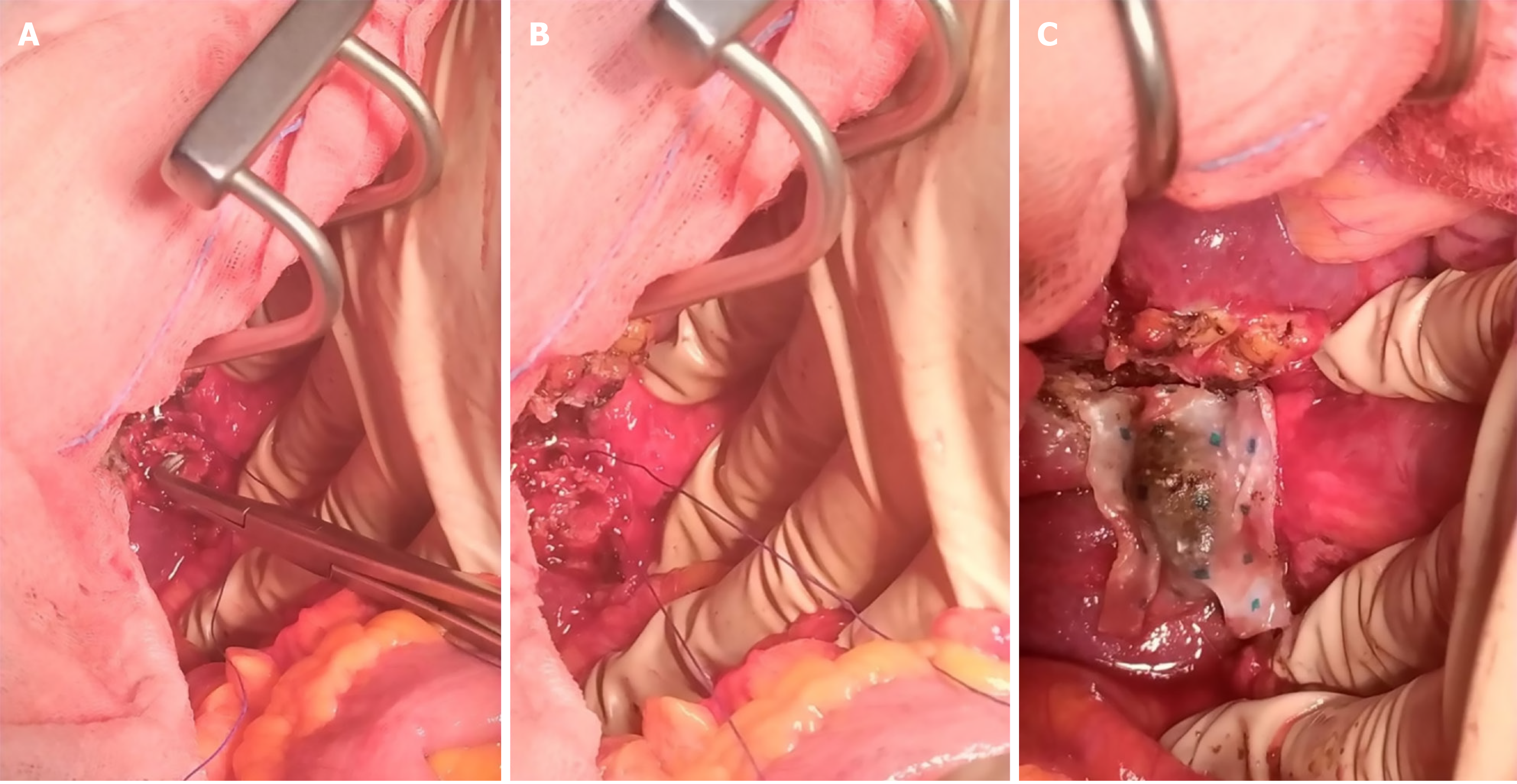
Figure 3 Reinforcement of the infundibular stump after subtotal cholecystectomy using Hemopatch®.
A: Infundibular stump following subtotal cholecystectomy for severe cholecystitis; B: Suture closure of the infundibular region; C: Placement of Hemopatch® over the suture line to reinforce the closure and reduce the risk of bile leakage.
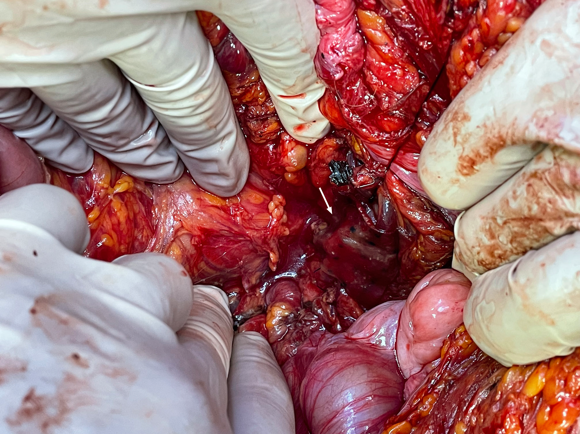
Figure 4 Reinforcement of the rectal stump after anterior rectal resection.
The rectal stump is covered with Hemopatch® to protect the suture line and potentially reduce the risk of postoperative leakage (white arrow).
- Citation: Zanatta M, Donati M. Hemopatch® as a sealant in anastomotic and suture leak prevention in abdominal emergency surgery: A single-center case series. World J Gastrointest Surg 2026; 18(9): 120497
- URL: https://www.wjgnet.com/1948-9366/full/v18/i9/120497.htm
- DOI: https://dx.doi.org/10.4240/wjgs.120497