
















Copyright: ©Author(s) 2026.
World J Gastrointest Surg. Aug 27, 2026; 18(8): 121170
Published online Aug 27, 2026. doi: 10.4240/wjgs.121170
Published online Aug 27, 2026. doi: 10.4240/wjgs.121170
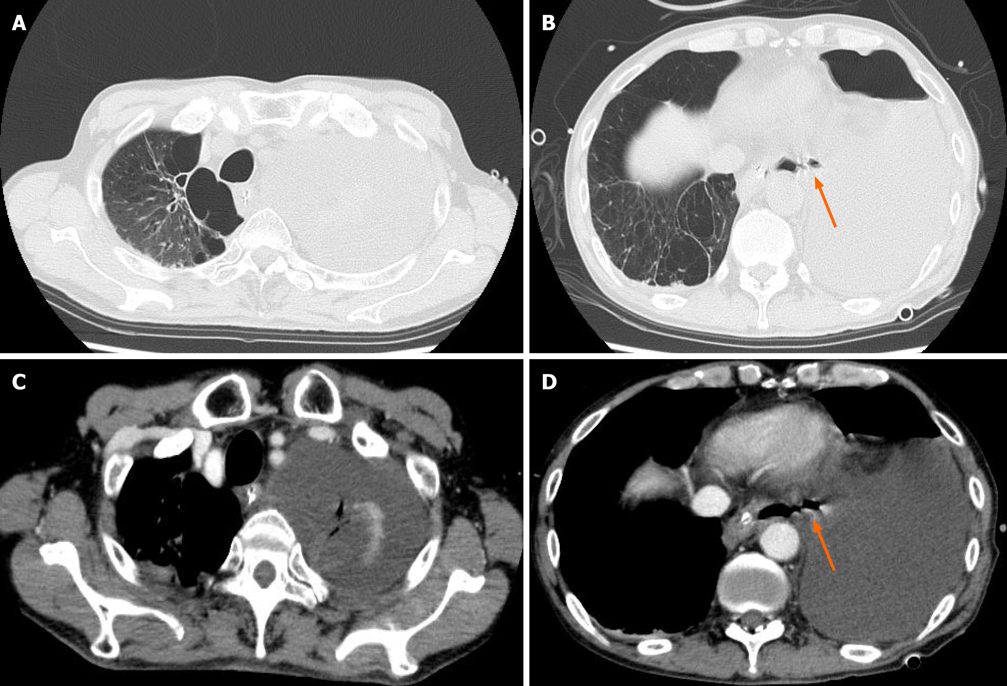
Figure 1 Contrast-enhanced computed tomography findings at the onset of spontaneous esophageal rupture.
A: A large amount of fluid was collected in the left thoracic cavity with left-lung collapse and rightward mediastinal shift, and a nasogastric tube for decompression was in place; B: Continuity between the lower esophageal lumen and the left pleural cavity suggestive of esophageal perforation; the ruptured esophageal wall is indicated by an arrow. No mediastinal or subcutaneous emphysema was observed; C: Extensive fluid was collected in the left thoracic cavity, with compression of the left lung; D: Suspected fistulous communication between the lower esophagus and the left pleural cavity; the ruptured esophageal wall is indicated by an arrow. Fluid accumulation with an air–fluid level was present in the left thoracic cavity.
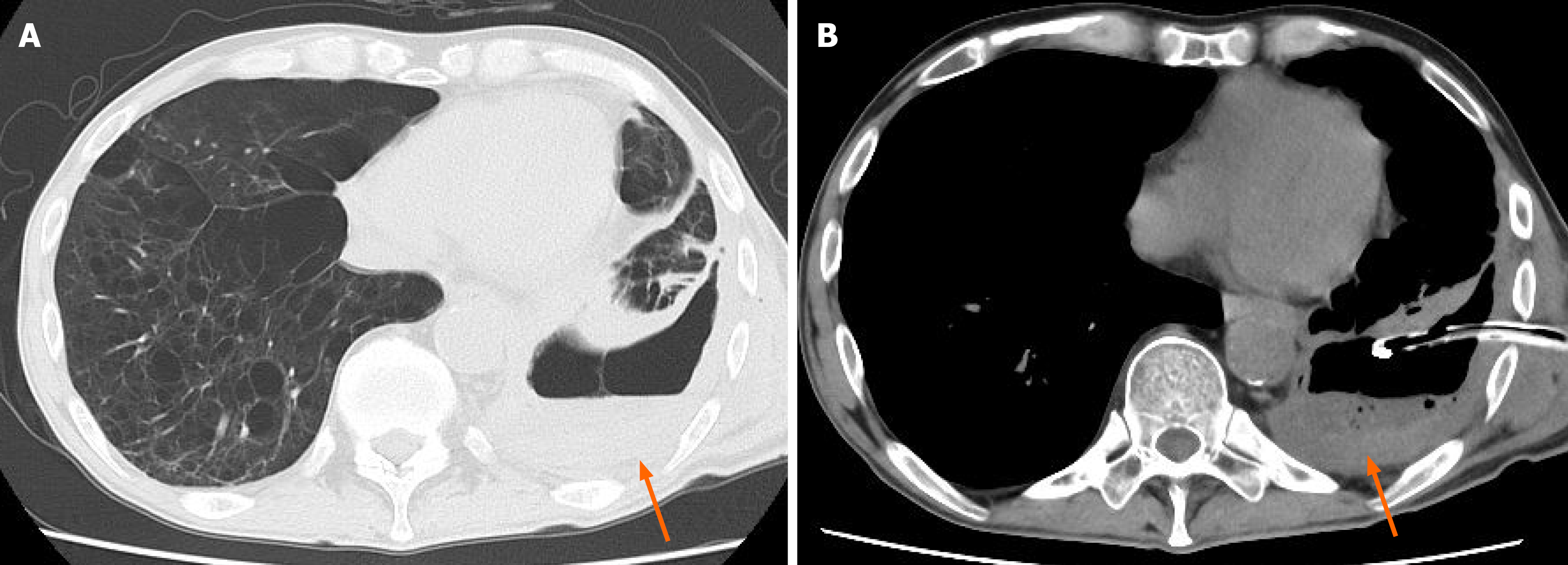
Figure 2 Plain computed tomography findings at the time of transfer to our hospital, 1 year after onset.
A: Recurrent fluid was collected with an air-fluid level in the left thoracic cavity and left-lung collapse; severe bilateral emphysematous changes were evident; B: Encapsulated fluid was collected with relatively high attenuation and intralesional free air in the left thoracic cavity, consistent with a pleural abscess; the abscess cavity in the thoracic cavity is indicated by an arrow. A chest tube was inserted into the abscess cavity for drainage, yielding saliva-like clear mucus and purulent discharge.
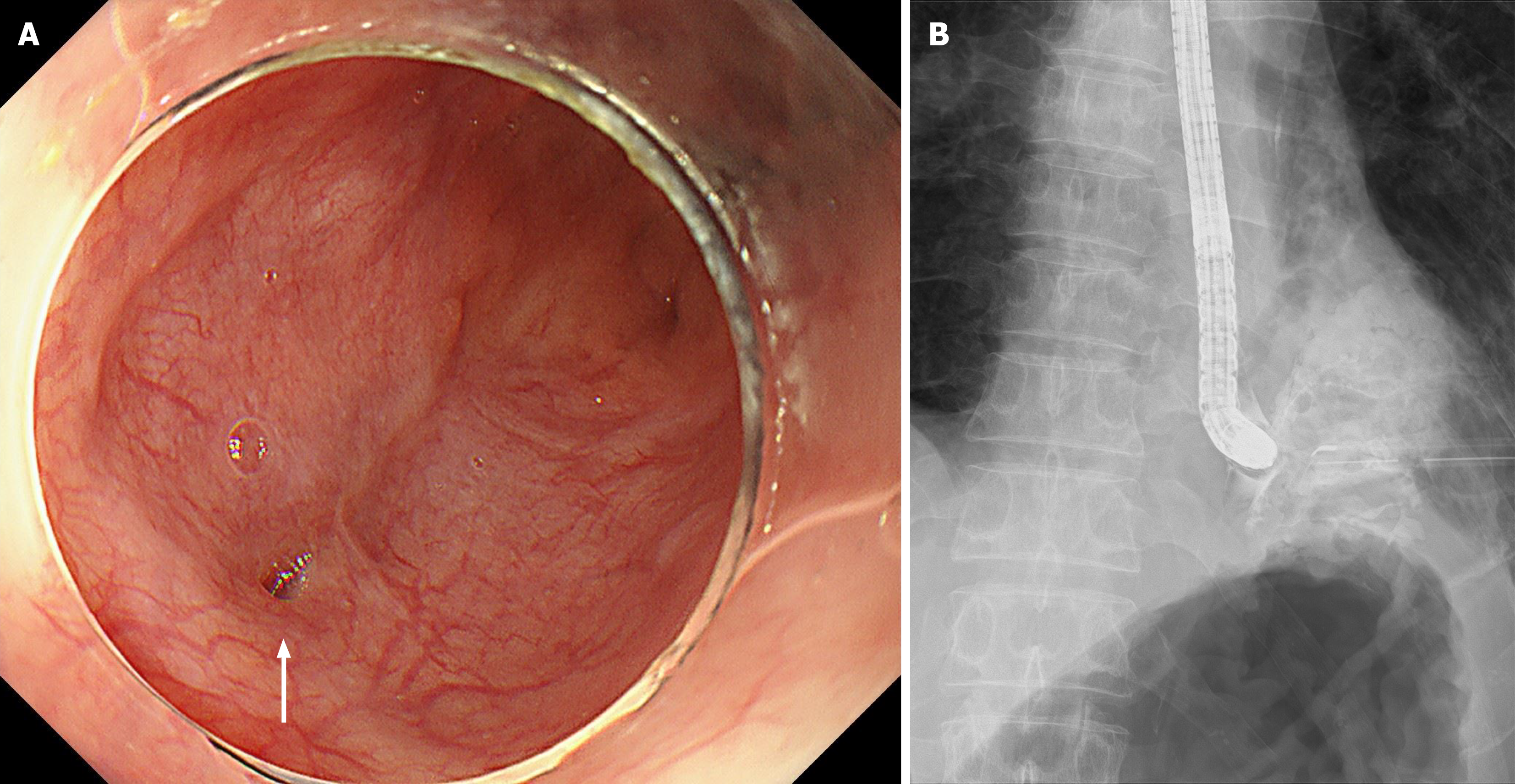
Figure 3 Endoscopic and contrast findings at the time of transfer to our hospital (1 year after onset).
A: Upper gastrointestinal endoscopy revealed a well-demarcated fistulous opening approximately 2 mm in diameter on the left wall just above the esophagogastric junction; the surrounding mucosa showed only mild erythema, without marked edema or necrotic changes; the esophagopleural fistula is indicated by an arrow; B: Contrast injection through a catheter inserted into the fistula demonstrated immediate flow of the contrast medium into the left pleural abscess cavity, confirming persistent communication of the esophagopleural fistula.
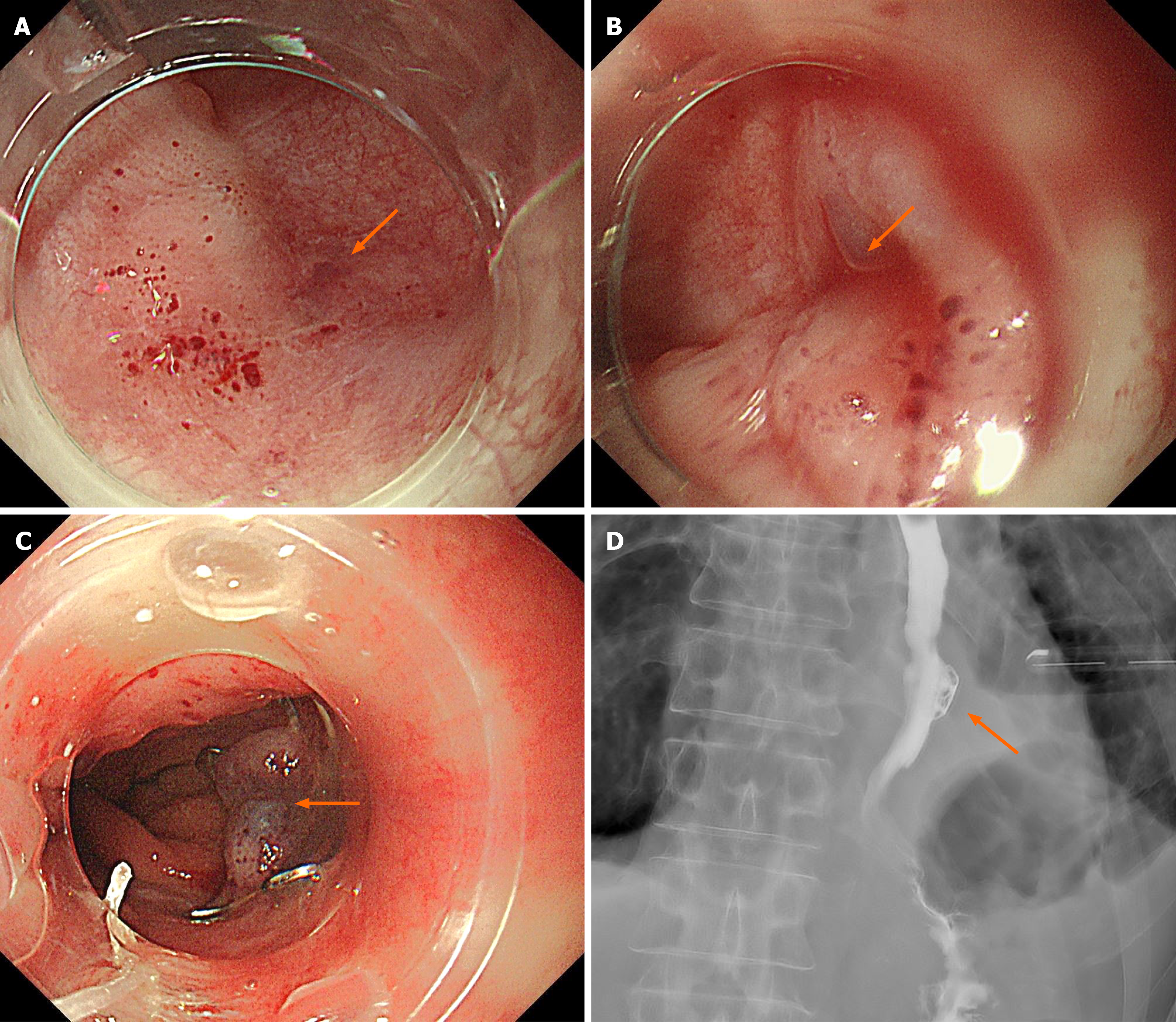
Figure 4 Endoscopic closure using an over-the-scope clip.
A: Endoscopic view of the 2-mm fistula located on the left wall just above the esophagogastric junction; B: The fistula and surrounding mucosa were suctioned into the over-the-scope clip (OTSC) cap to achieve adequate tissue capture; C: The OTSC was deployed after confirmation of sufficient tissue inversion into the cap, achieving full-thickness closure; D: Immediate water-soluble esophagography after OTSC placement demonstrated no visualization of the fistula and no leakage into the left pleural cavity; additional clips were not required; the esophagopleural fistula is indicated by an arrow.
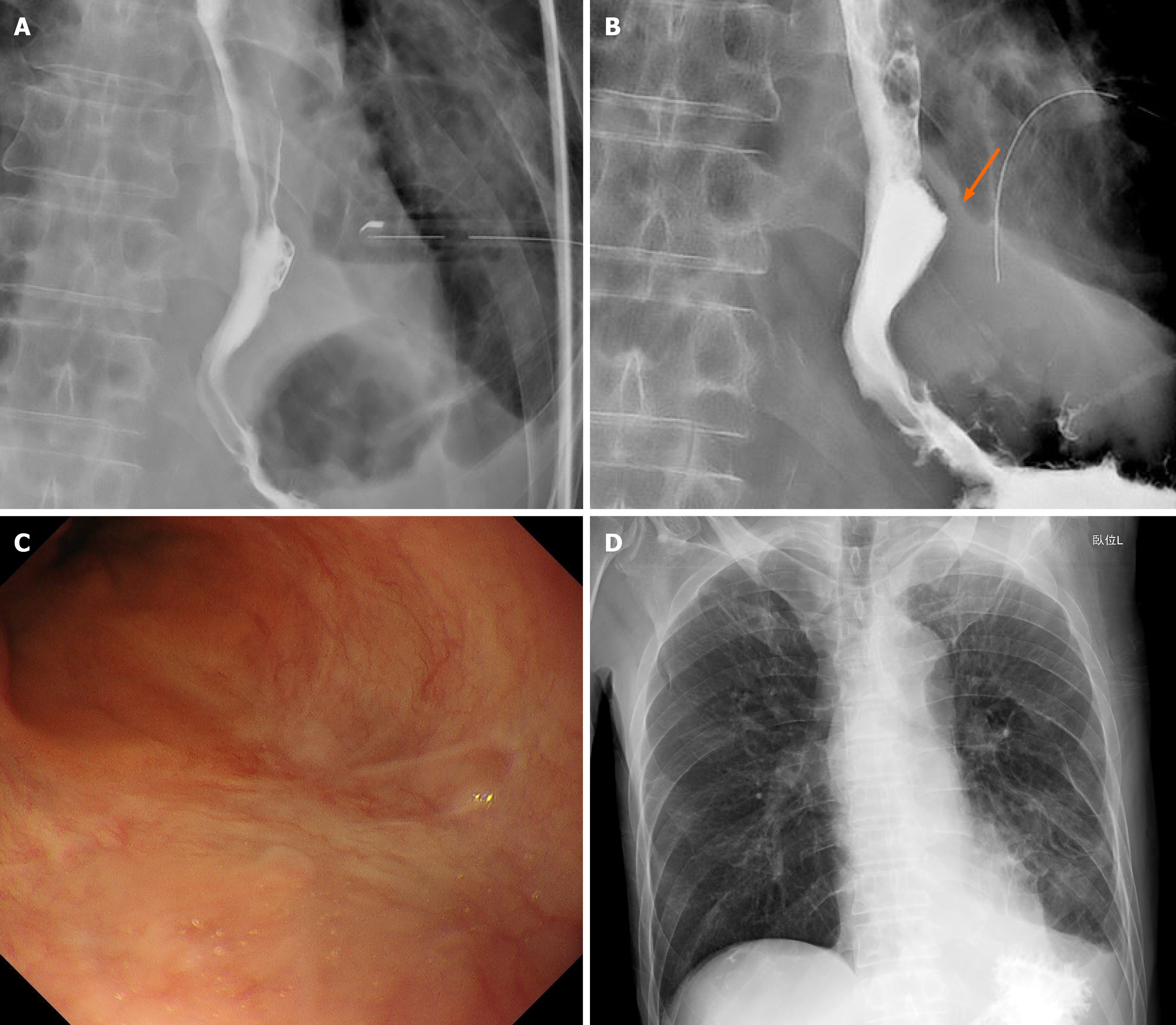
Figure 5 Follow-up imaging after over-the-scope clip placement.
A: Water-soluble contrast esophagography performed 14 days after over-the-scope clip (OTSC) placement showed no leakage into the thoracic cavity; B: Water-soluble contrast esophagography performed 27 days after OTSC placement showed no leakage despite spontaneous OTSC dislodgement; the previously existing fistula site, now closed, is indicated by an arrow; C: Follow-up endoscopic view confirming clip dislodgement and progressive mucosal healing at the previous fistula site without recurrence; D: Chest radiograph showing improved radiolucency in the left lower-lung field, corresponding to the resolution of empyema.
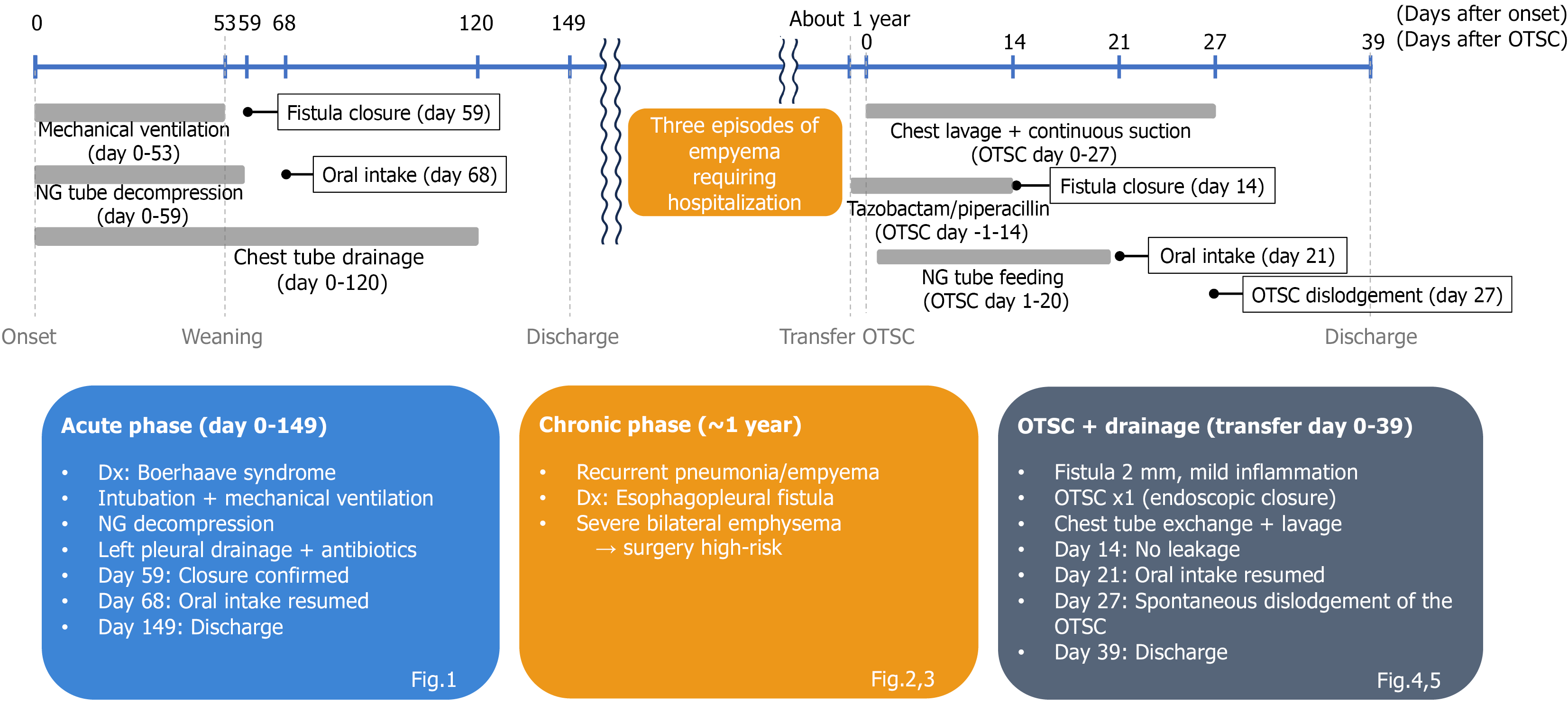
Figure 6 Clinical course.
Timeline of the patient’s clinical course from onset of spontaneous esophageal rupture to discharge after over-the-scope clip (OTSC) treatment. The upper axis indicates the days after onset, while the lower axis indicates the days after OTSC placement. Mechanical ventilation was required from day 0 to day 53. Fistula closure was confirmed on day 59, and the patient was discharged on day 149. Approximately 1 year later, an esophagopleural fistula was diagnosed and treated with OTSC and continuous drainage. Contrast esophagography confirmed closure on day 14 after OTSC placement. Spontaneous clip dislodgement occurred on day 27 after OTSC placement without recurrence, and the patient was discharged on day 39 after OTSC placement. OTSC: Over-the-scope clip.
- Citation: Takahashi N, Baba S, Toya Y, Nikai H, Fujisawa R, Endo F, Umemura A, Iwaya T, Nitta H, Sasaki A. Curative endoscopic closure of a chronic esophagopleural fistula after Boerhaave syndrome using an over-the-scope clip: A case report. World J Gastrointest Surg 2026; 18(8): 121170
- URL: https://www.wjgnet.com/1948-9366/full/v18/i8/121170.htm
- DOI: https://dx.doi.org/10.4240/wjgs.121170