
















Copyright: ©Author(s) 2026.
World J Gastrointest Surg. Aug 27, 2026; 18(8): 121147
Published online Aug 27, 2026. doi: 10.4240/wjgs.121147
Published online Aug 27, 2026. doi: 10.4240/wjgs.121147
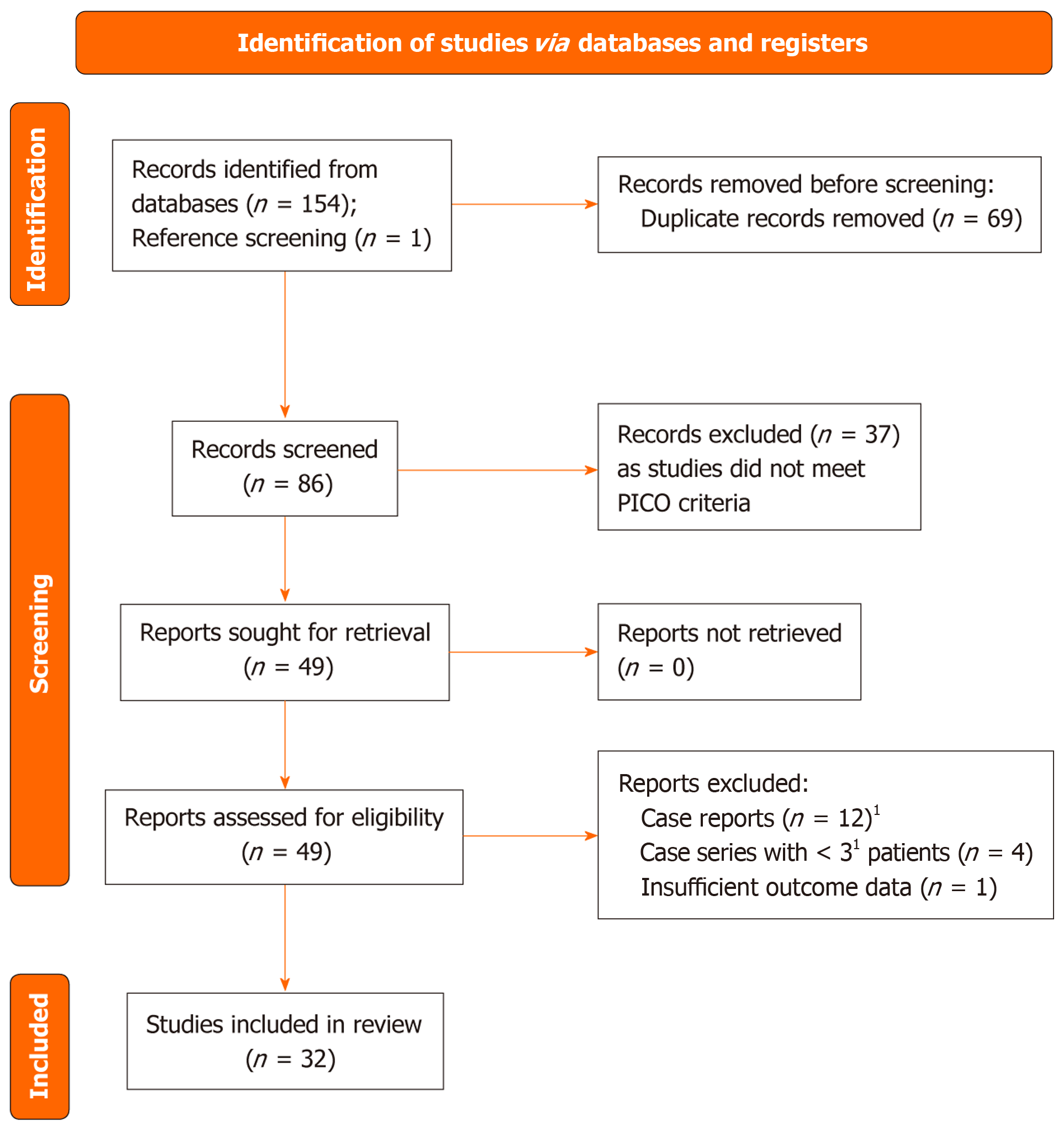
Figure 1 Flow diagram of study selection.
The diagram summarises the identification, screening, exclusion, and inclusion of studies used in the narrative review and evidence tables. 1Selected case reports were included in the descriptive discussion when they were innovative or of historical significance. PICO: Patient population, intervention, comparison, and outcome.
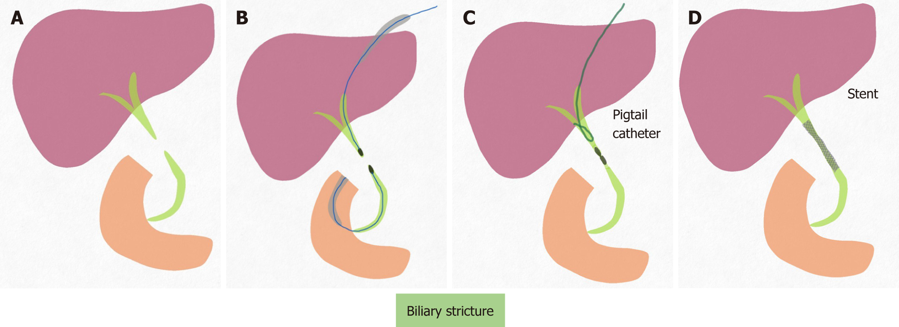
Figure 2 Magnetic compression anastomosis for biliary stricture.
A: Post-operative or post-transplant biliary stricture; B: Delivery of Proximal and distal magnet via percutaneous transhepatic catheter and endoscopy; C: Magnet approximated and stricture being negotiated by wire; D: Once neo-fistula formed and wire negotiated, stent is placed to maintain the patency of the fistula.
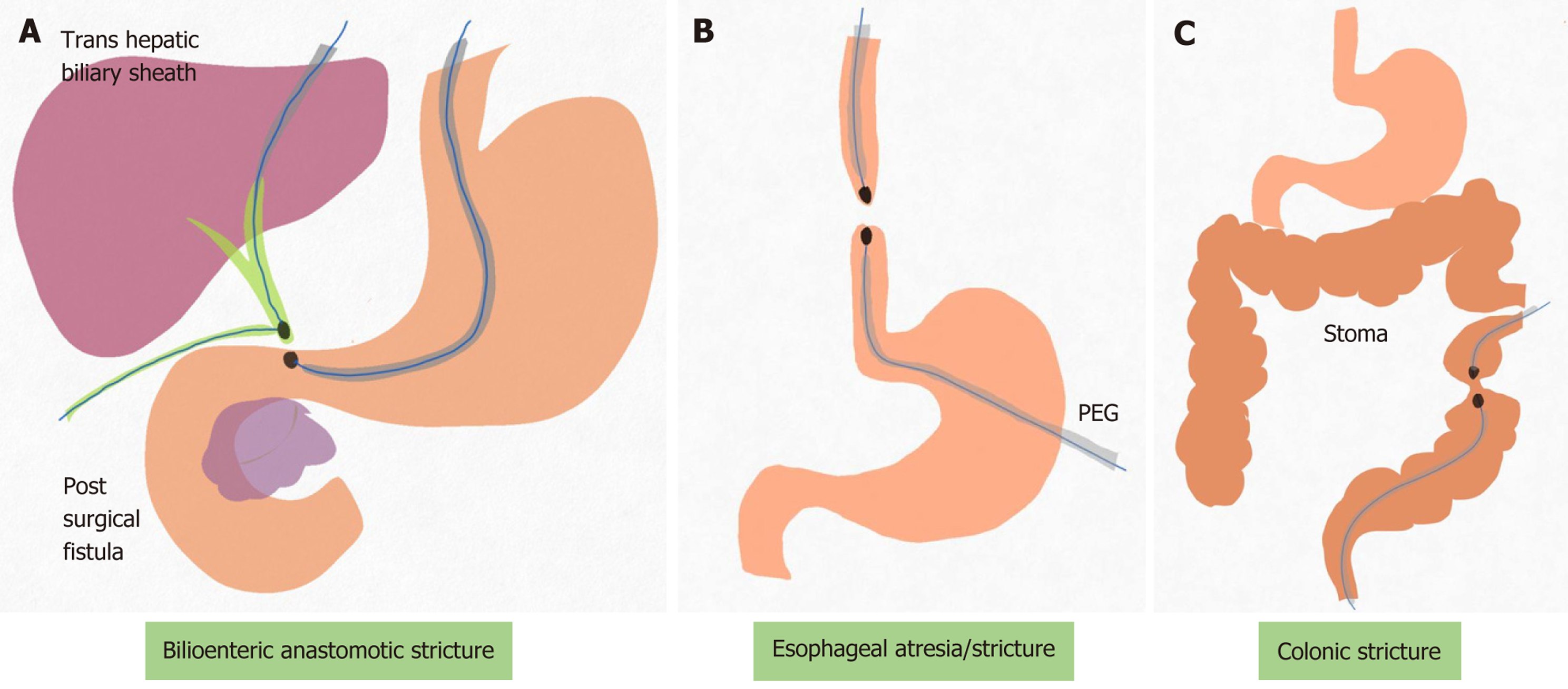
Figure 3 Representative access routes for magnetic compression anastomosis in gastrointestinal and biliary disease.
A: Bilioenteric anastomotic stricture approached through transhepatic and enteroscopic routes; B: Esophageal anastomotic stricture approached through oral endoscopy and gastrostomy access; C: Colorectal stricture approached through colonoscopic and distal luminal access. PEG: Percutaneous endoscopic gastrostomy.
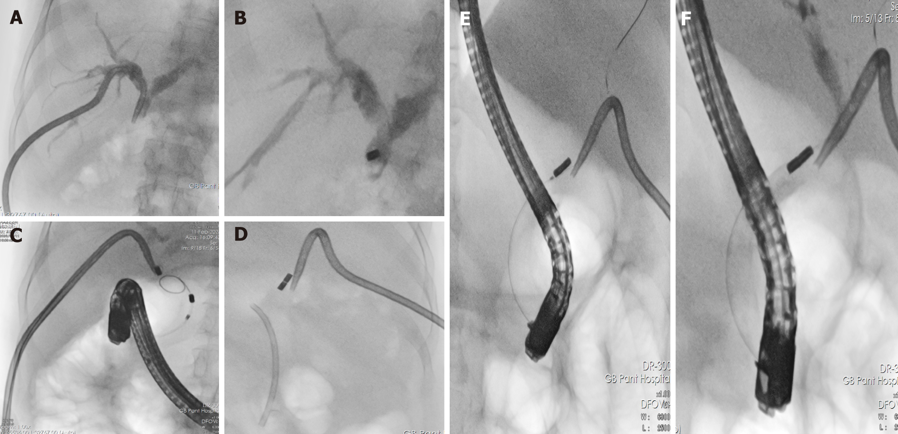
Figure 4 Magnetic compression anastomosis for postcholecystectomy bile duct injury stricture.
A: Percutaneous transhepatic cholangiogram showing complete cut off of common bile duct; B: Parent magnet being deployed by percutaneous trans hepatic route; C: Daughter magnet being delivered via endoscopic retrograde cholangiographic route; D: Both the magnet were kept in place by stents; E: After 10 days, magnets approximated and wire negotiated into intrahepatic biliary radicle via neo fistula; F: Cholangiogram demonstrating resolution of stricture and neo fistula.
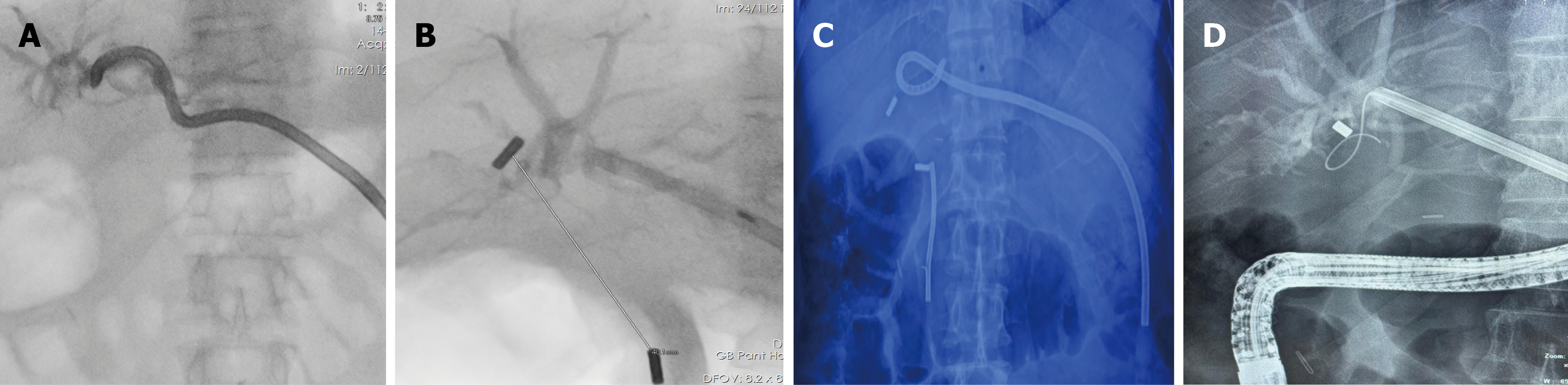
Figure 5 Magnet migration and failed magnetic compression anastomosis.
A: Percutaneous cholangiogram showing complete cut of the bile duct; B: Both parent and daughter magnets deployed, however, the distance between them is long (4 cm); C: Serial radiograph showed no anastomosis formation; D: Distal magnet was retrieved endoscopically; however, cholangioscopic retrieval of the magnet failed, and the patient underwent surgery.
- Citation: Reddy RT, Bagrodia A, Mehta K, Vaithiyam V, Mohan SL, Sirohi N, Dalal A, Srivastava S, Sachdeva S. Magnetic compression anastomosis in gastrointestinal and biliary disease: Techniques and clinical applications. World J Gastrointest Surg 2026; 18(8): 121147
- URL: https://www.wjgnet.com/1948-9366/full/v18/i8/121147.htm
- DOI: https://dx.doi.org/10.4240/wjgs.121147