
















Copyright: ©Author(s) 2026.
World J Gastrointest Surg. Aug 27, 2026; 18(8): 120441
Published online Aug 27, 2026. doi: 10.4240/wjgs.120441
Published online Aug 27, 2026. doi: 10.4240/wjgs.120441
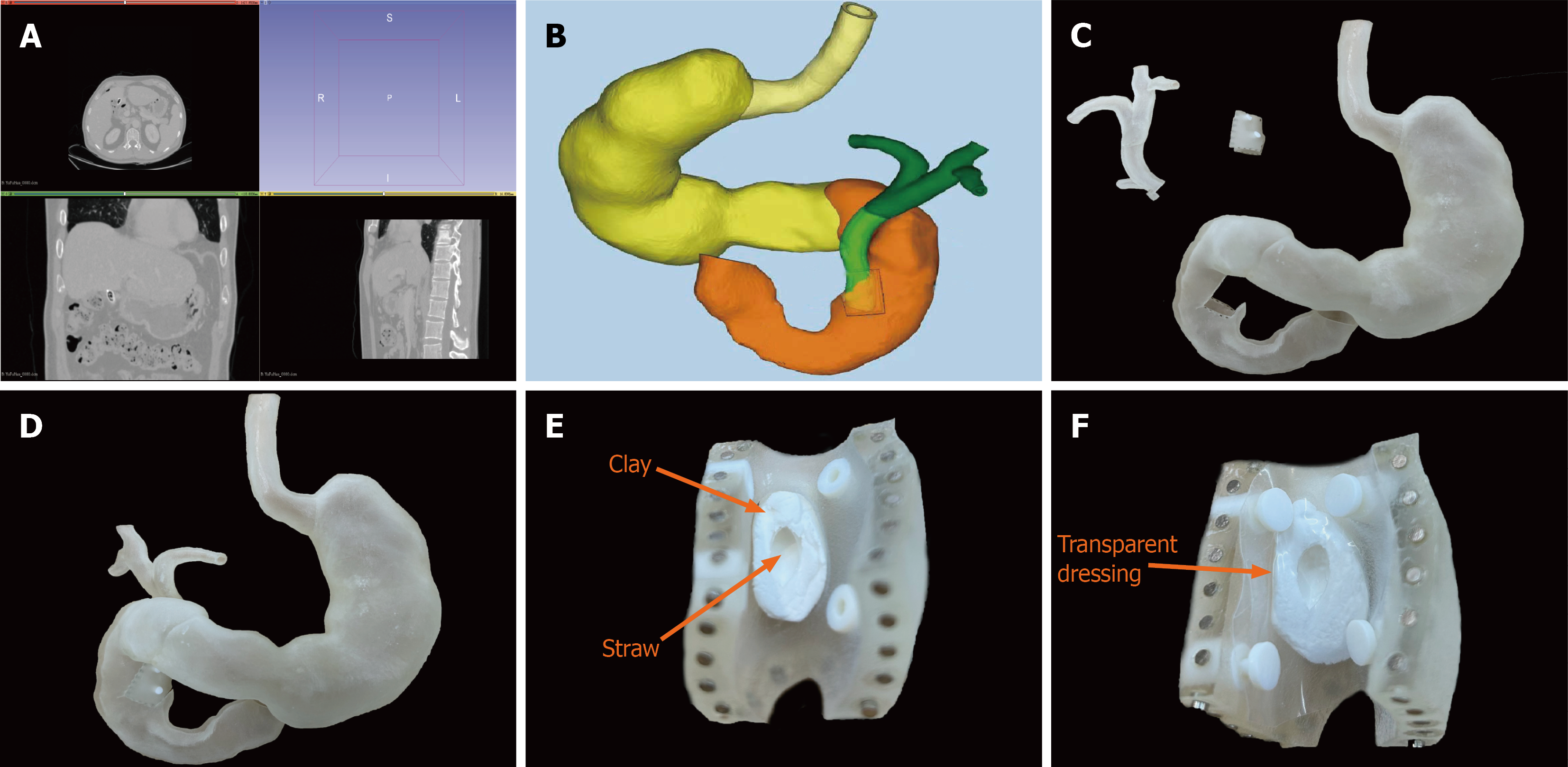
Figure 1 Construction of a three-dimensional-printed model based on computed tomography data.
A: Computed tomography scan data of endoscopic retrograde cholangiopancreatography patients before surgery; B: Construction of the three-dimensional (3D) simulation model; C: Division of the model into the gastroduodenal, common bile duct, and duodenal connection sections; D: 3D-printed model after reconstruction; E and F: Design of the duodenal papilla connection section.
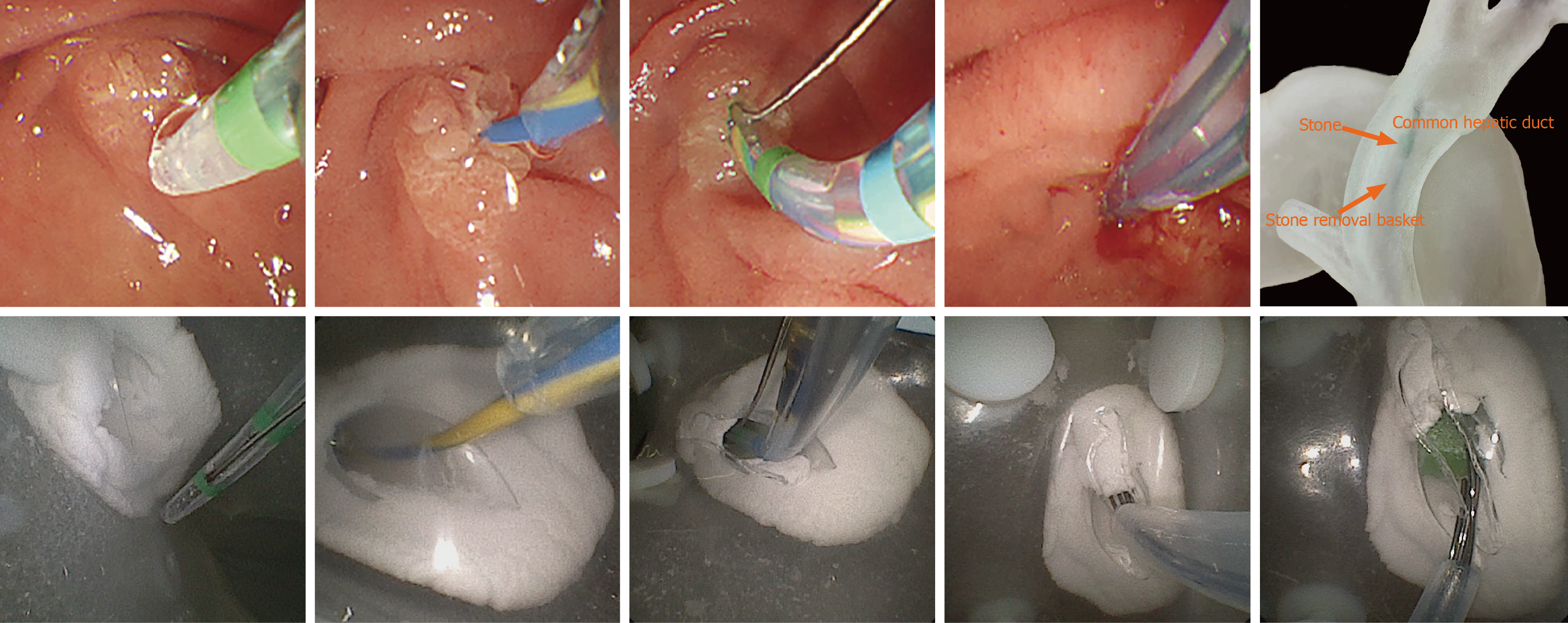
Figure 2 Simulation of the endoscopic retrograde cholangiopancreatography surgical process.
Comparison of practical (top) and model operation (bottom). From left to right: Duodenal papilla identification; Guidewire insertion; Papillary incision; Stone basket insertion; Stone extraction.
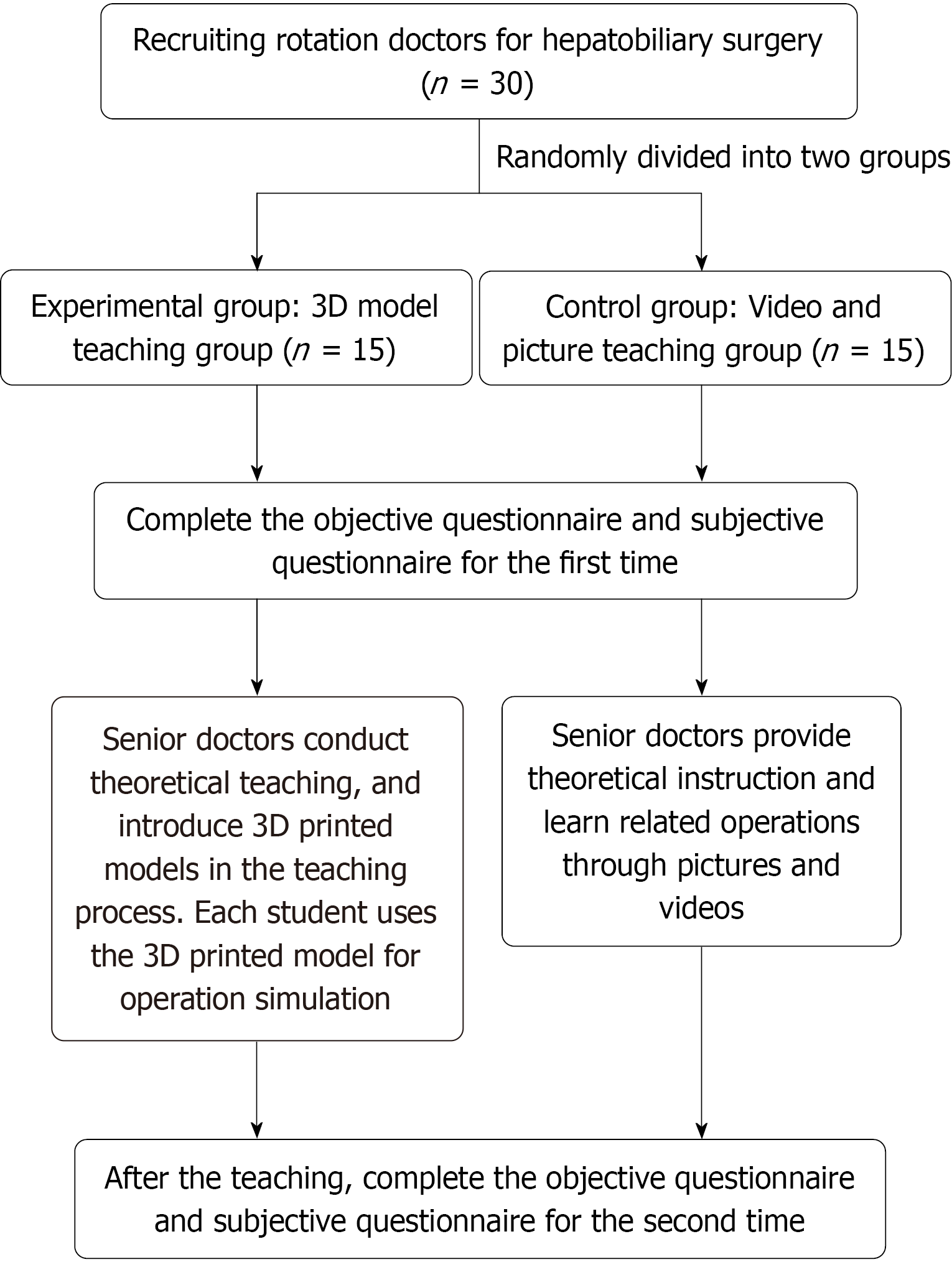
Figure 3 Diagram of the teaching model.
3D: Three-dimensional.
- Citation: Zhou WJ, Qin JW, Li C, Li XF. Application of a three-dimensional-printed endoscopic retrograde cholangiopancreatography simulation operation model in surgical training and teaching. World J Gastrointest Surg 2026; 18(8): 120441
- URL: https://www.wjgnet.com/1948-9366/full/v18/i8/120441.htm
- DOI: https://dx.doi.org/10.4240/wjgs.120441