
















Copyright: ©Author(s) 2026.
World J Gastrointest Surg. Aug 27, 2026; 18(8): 120360
Published online Aug 27, 2026. doi: 10.4240/wjgs.120360
Published online Aug 27, 2026. doi: 10.4240/wjgs.120360
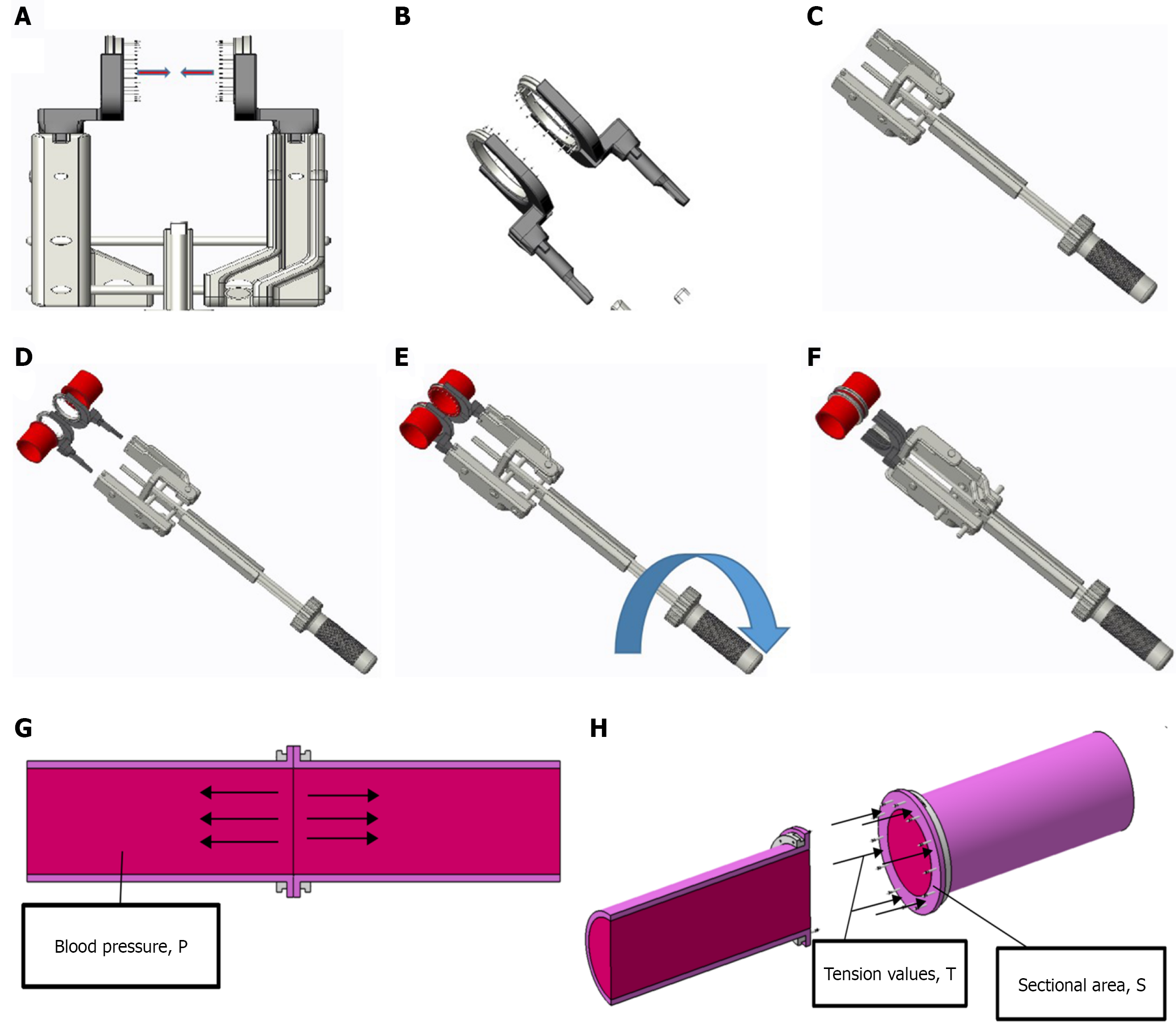
Figure 1 Sequential steps of microvascular anastomotic coupler application.
A: Align the transected portal vein ends and select the coupler size with a vascular gauge (outer diameter matched to vessel caliber); B: Open the coupler and expose the barbed pins; C: Evert each vein edge and seat the wall evenly onto the pins, ensuring full thickness capture without intimal folding; D: Repeat pinning on the contralateral stump with circumferential, evenly spaced engagement; E: Approximate the rings and lock using the mechanical rotation tool until the audible/visual stop is reached; F: Inspect for uniform apposition, absence of gaps/bleeding, and confirm lumen patency; G and H: Schematics showing pressure and tension distribution after closure, highlighting circumferential load sharing and anti-slippage by the barbed pins. P: Pressure; T: Tension; S: Sectional.
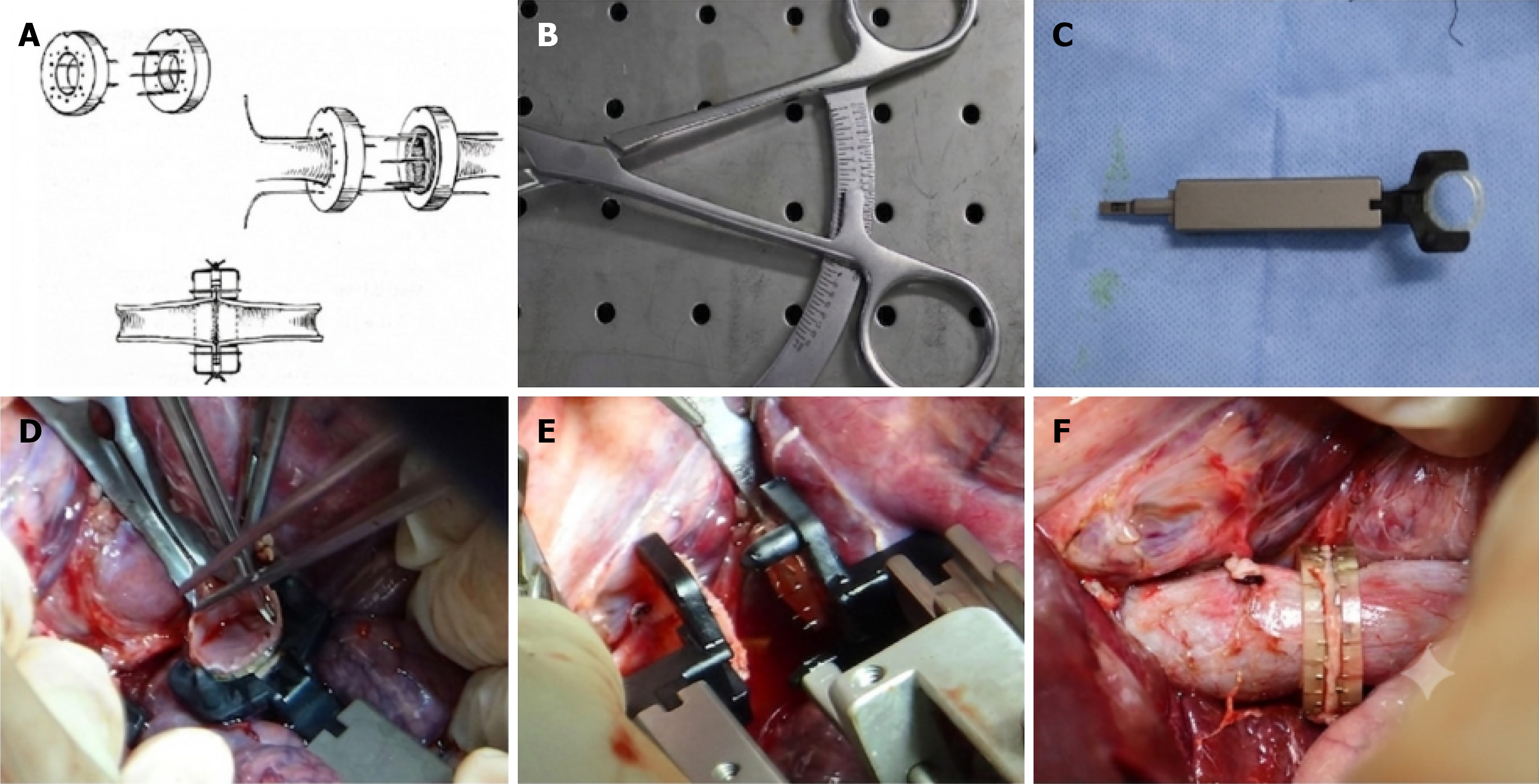
Figure 2 Device overview and surgical application for portal vein reconstruction.
A: Schematic illustration of the microvascular anastomotic coupler (KingSung Medical; NMPA Reg. No. 20233020825) and its alignment mechanism; B: Vascular gauge for diameter measurement; C: The coupler device; D: Intraoperative preparation of the portal vein ends; E: Alignment and closure of the vessel ends using the rotation tool; F: Completed anastomosis showing a secure, uniform connection.
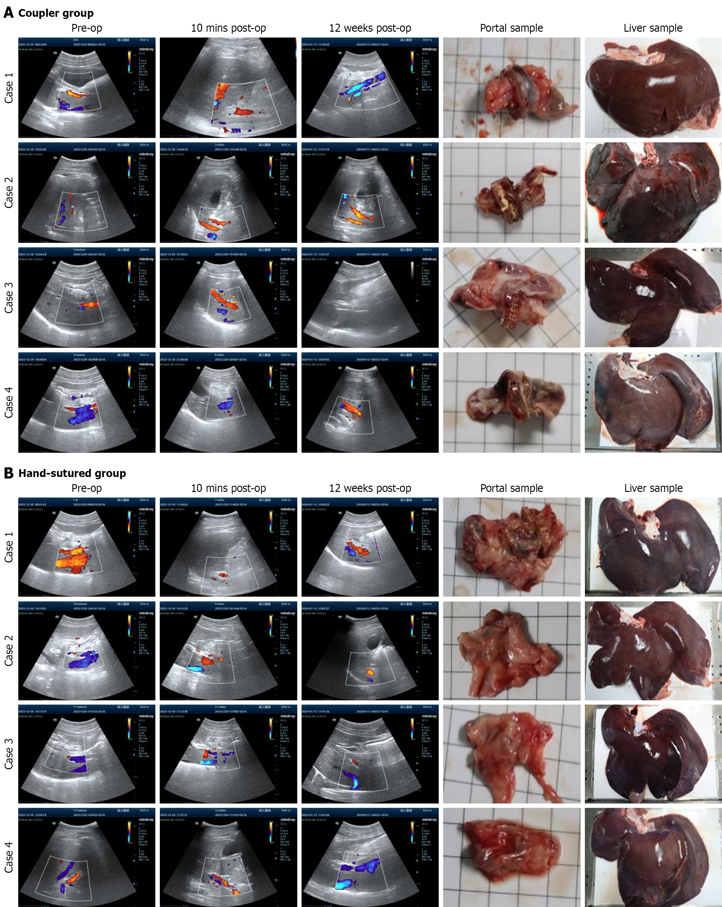
Figure 3 Doppler ultrasound and gross specimen evaluation.
A: Doppler ultrasound and specimen evaluation of the coupler group. Columns 1-3: Doppler ultrasound images of portal vein velocity at pre-operation, 10 minutes post-operation, and 12 weeks post-operation for four representative cases. These images demonstrate stable velocity and patency. Column 4: Portal vein samples excised at 12 weeks, showing uniform healing and intact structures. Column 5: Liver specimens from the same animals, showing no visible ischemic damage; B: Doppler ultrasound and specimen evaluation of the hand-sutured group. Columns 1-3: Doppler ultrasound images at the same time points for four representative cases. Variable velocity and signs of stenosis or reduced patency are observed in some cases at 12 weeks. Column 4: Portal vein samples, showing occasional irregular healing and fibrosis. Column 5: Liver specimens, with some cases showing mild ischemic changes.
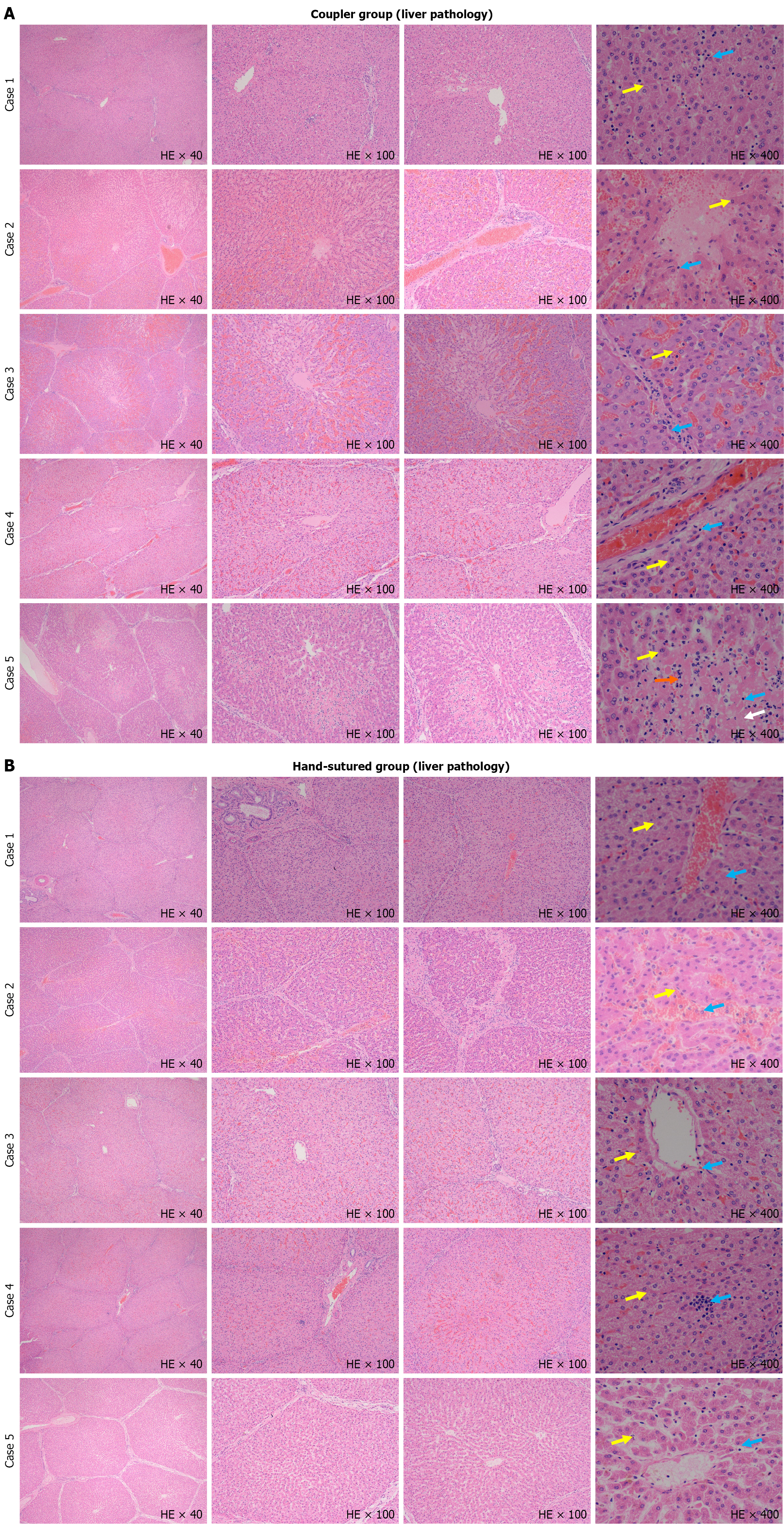
Figure 4 Histological analysis of liver tissue in the coupler group/hand-sutured group.
A: Histological analysis of liver tissue in the coupler group (hematoxylin and eosin staining). Representative images from five cases at 40 ×, 100 ×, and 400 ×. 40 ×/100 ×: Lobular architecture is preserved without significant inflammation, fibrosis, or necrosis. 400 ×: High magnification views show intact hepatocytes (yellow arrows), scattered lymphocytes (blue arrows), small necrotic areas (white arrows), and occasional polymorphonuclear cells (orange arrows), indicating minimal injury; B: Histological analysis of liver tissue in the hand-sutured group (hematoxylin and eosin staining). Representative images from five cases at 40 ×, 100 ×, and 400 ×. 40 ×/100 ×: Mild disorganization of lobular structure, focal necrosis, and inflammatory cell infiltration are seen in some cases. 400 ×: Intact hepatocytes (yellow arrows) and lymphocytes (blue arrows) are present, but overall inflammatory burden is higher.
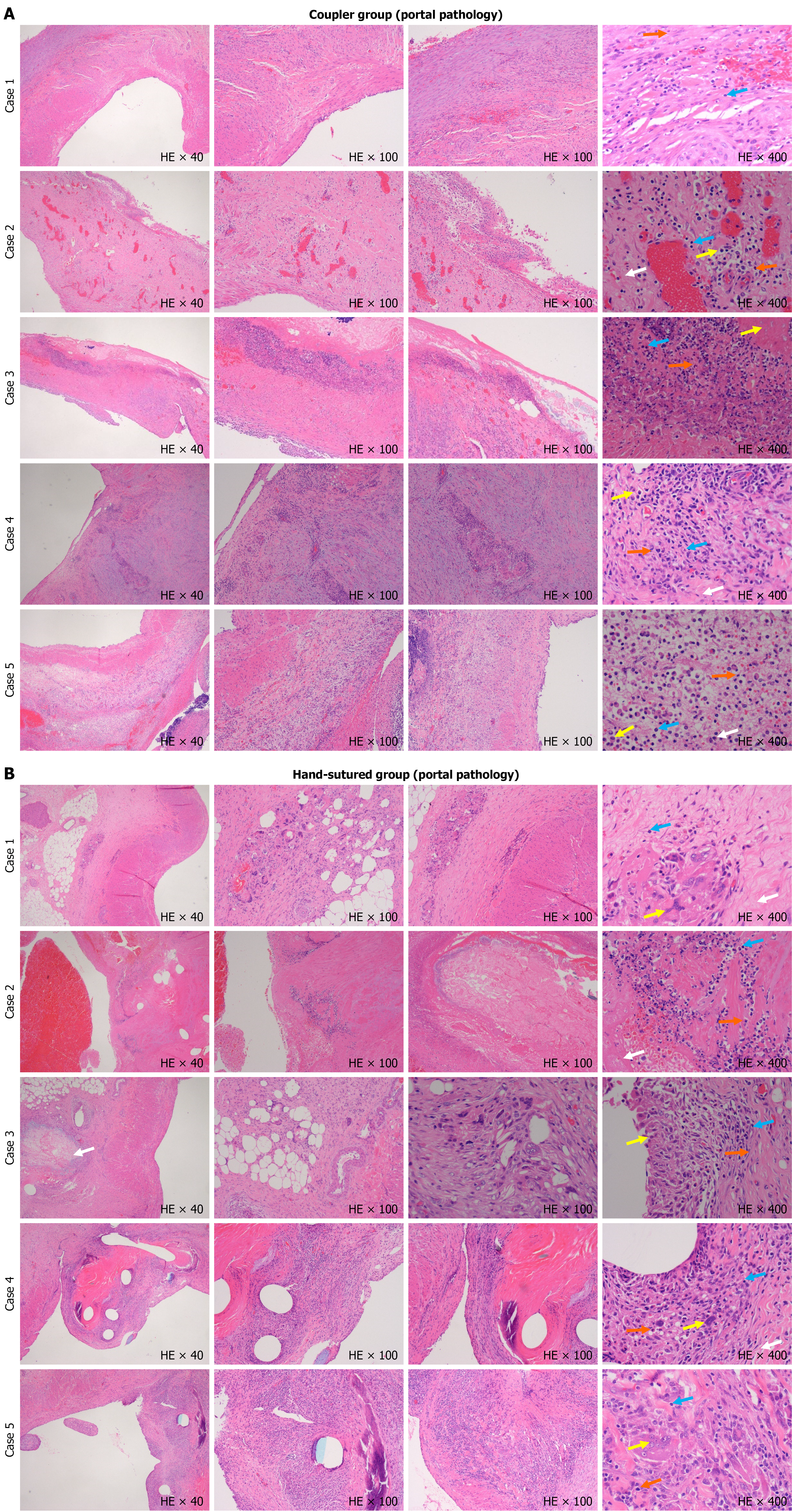
Figure 5 Histological analysis of portal vein tissue in the coupler group/hand-sutured group.
A: Histological analysis of portal vein tissue in the coupler group (hematoxylin and eosin staining). Representative images from five cases at 40 ×, 100 ×, and 400 ×. 40 ×/100 ×: The portal vein exhibits intact structure with minimal inflammation and no significant necrosis. Vessel walls are well preserved. 400 ×: Scattered macrophages (yellow arrows), lymphocytes (blue arrows), and rare plasma cells (orange arrows) are seen, with only small necrotic areas (white arrows); B: Histological analysis of portal vein tissue in the hand-sutured group (hematoxylin and eosin staining). Representative images from five cases at 40 ×, 100 ×, and 400 ×. 40 ×/100 ×: Structural disorganization, inflammatory cell infiltration, and focal necrosis are evident. Vessel walls show irregular thickening and fibrosis. 400 ×: Increased macrophages (yellow arrows), lymphocytes (blue arrows), plasma cells (orange arrows), and significant necrotic tissue (white arrows) are present, indicating higher inflammation and tissue injury.
- Citation: Li JJ, Yang J, Xie Y, Shang JM, Li J, Chen CY, Liu LN, Zhang ZY, Pan BH, Jiang WT. Sutureless portal vein reconstruction in porcine orthotopic liver transplantation using a microvascular coupler: A randomized study with artificial intelligence-assisted histopathology. World J Gastrointest Surg 2026; 18(8): 120360
- URL: https://www.wjgnet.com/1948-9366/full/v18/i8/120360.htm
- DOI: https://dx.doi.org/10.4240/wjgs.120360