
















Copyright: ©Author(s) 2026.
World J Gastrointest Surg. Jul 27, 2026; 18(7): 121328
Published online Jul 27, 2026. doi: 10.4240/wjgs.v18.i7.121328
Published online Jul 27, 2026. doi: 10.4240/wjgs.v18.i7.121328
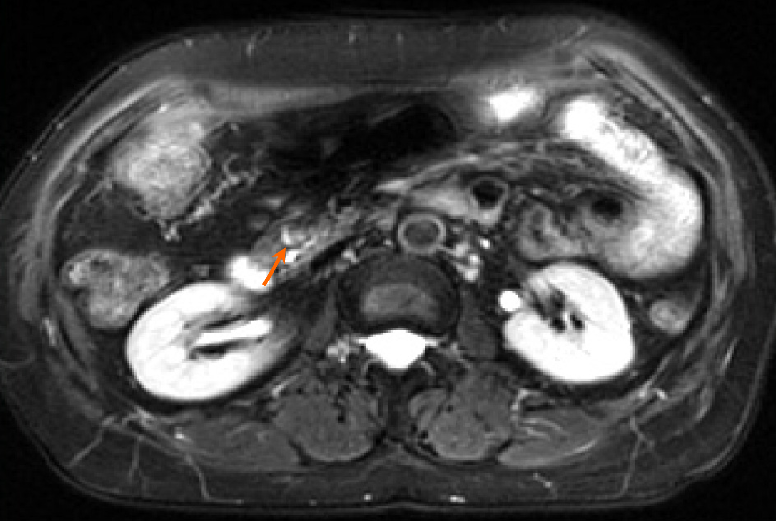
Figure 1 An enhanced abdominal magnetic resonance imaging suggested a possible intraductal papillary mucinous neoplasm.
Orange arrow: Intraductal papillary mucinous neoplasm.
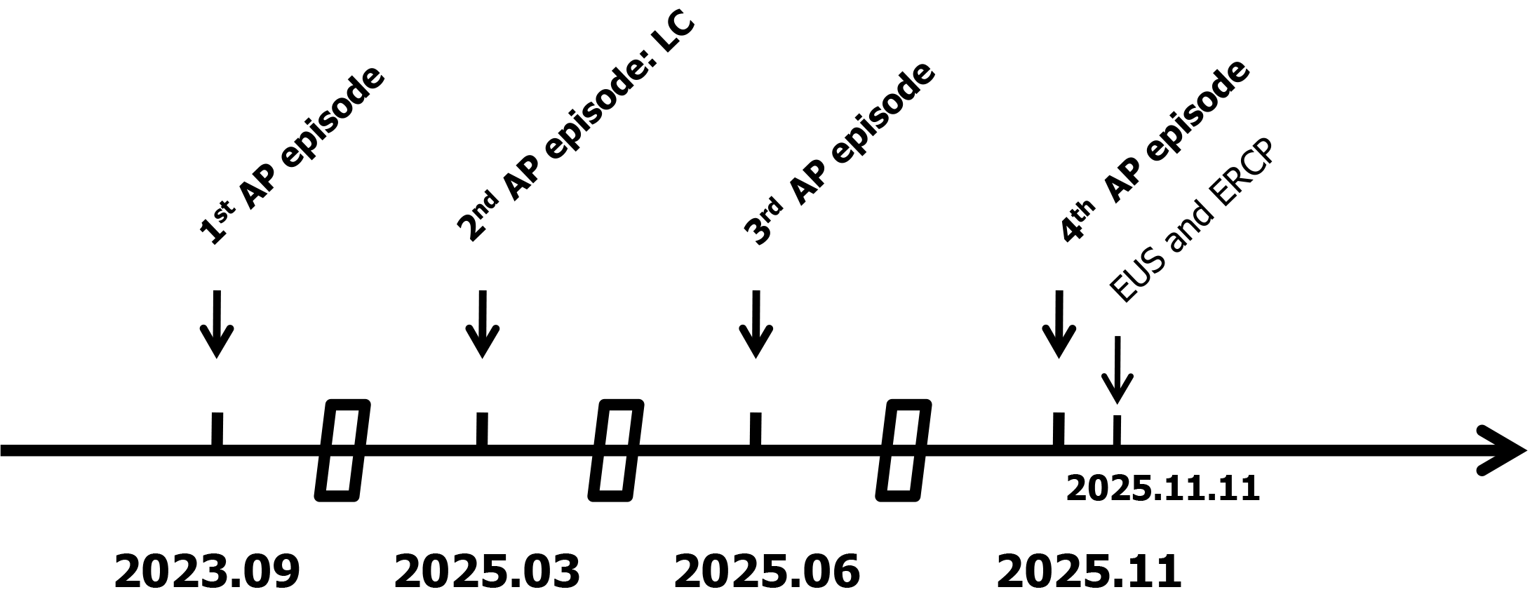
Figure 2 Timeline of the patient’s previous pancreatitis history.
AP: Acute pancreatitis; LC: Laparoscopic cholecystectomy; EUS: Endoscopic ultrasound.
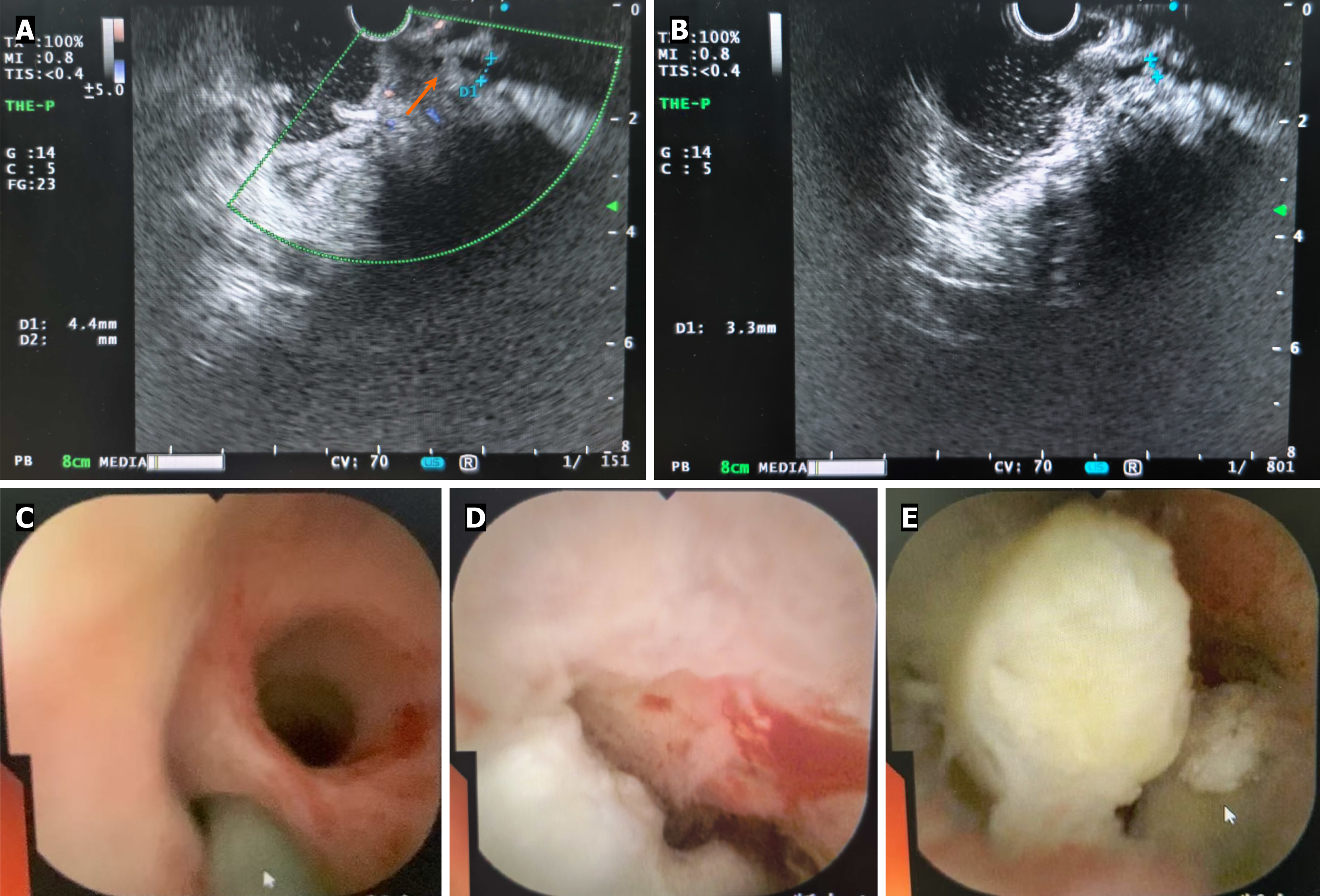
Figure 3 Representative images of branch-duct type intraductal papillary mucinous neoplasm and its mucous plug.
A and B: Endoscopic ultrasound revealed a distinct, roundish and hyperechoic structure causing the distal pancreatic duct to expand; C-E: Pancreatoscopy visualized multiple mucus plugs.
- Citation: Han X, Guo XY, Huang YS, Yu G, Lu J, Wan R, Yang LJ, Chen CY. Recurrent acute pancreatitis due to a subtle branch-duct type intraductal papillary mucinous neoplasm: A case report. World J Gastrointest Surg 2026; 18(7): 121328
- URL: https://www.wjgnet.com/1948-9366/full/v18/i7/121328.htm
- DOI: https://dx.doi.org/10.4240/wjgs.v18.i7.121328