
















Copyright: ©Author(s) 2026.
World J Gastrointest Surg. Jul 27, 2026; 18(7): 120929
Published online Jul 27, 2026. doi: 10.4240/wjgs.v18.i7.120929
Published online Jul 27, 2026. doi: 10.4240/wjgs.v18.i7.120929
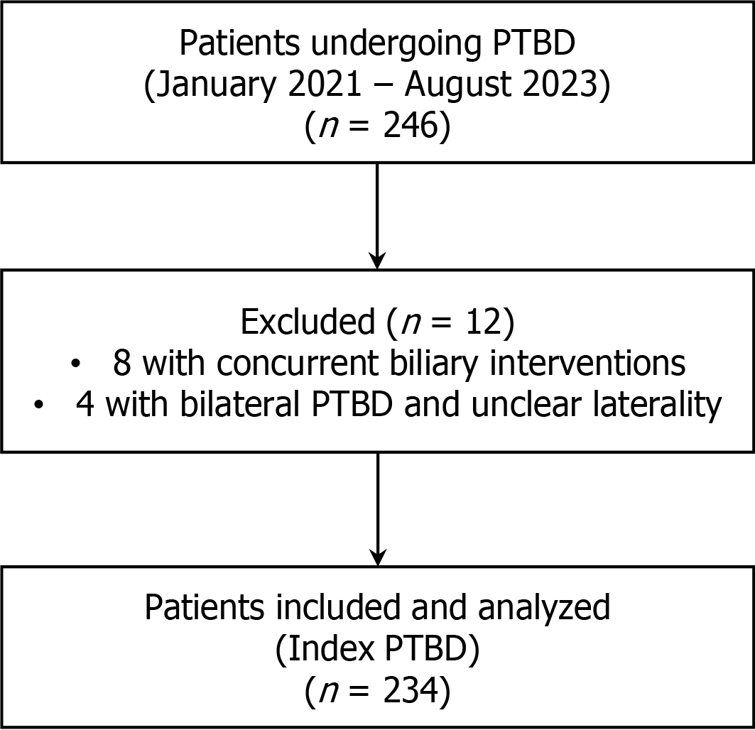
Figure 1 Study flow diagram of patient selection and inclusion for analysis.
PTBD: Percutaneous transhepatic biliary drainage.
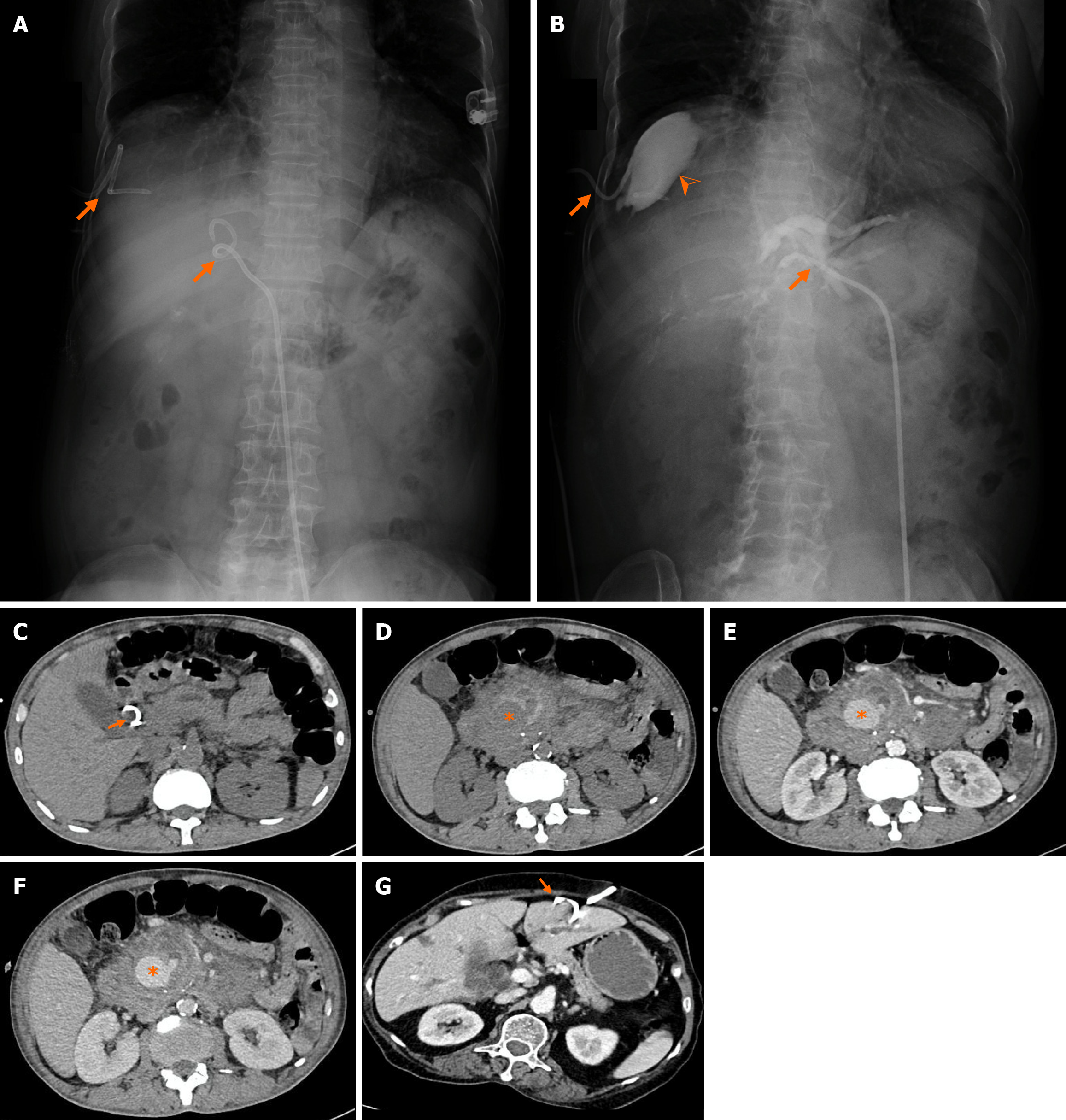
Figure 2 Procedure-related complications.
A and B: Catheter dislodgement on cholangiogram in a case of biliary obstruction due to a Klatskin tumor with bilateral biliary drainage. Image A is a cholangiogram before contrast injection, with orange arrows indicating the positions of the left and right biliary drainage catheters. Image B is a cholangiogram after contrast injection: The left catheter remains in the correct position, and contrast fills the left intrahepatic ducts up to the hepatic hilum. However, the right catheter has become dislodged from the biliary tract, resulting in contrast extravasation into the right subphrenic space (orange arrowhead), with no visualization of the right intrahepatic bile ducts; C-F: Pseudoaneurysm images on axial, arterial phase abdominal multi-slice computed tomography scan in a case of biliary obstruction due to a pancreatic head tumor with right hepatic duct drainage. Image C (non-contrast phase) shows the distal tip of the catheter located within the common bile duct (orange arrow). Images D (non-contrast), E (arterial phase), and F (venous phase) at the level of the pancreatic head reveal a large retroperitoneal hematoma containing a pseudoaneurysm, without evidence of contrast extravasation. The lesion appears as a low-attenuation area on non-contrast phase, becomes hyperattenuating in the arterial phase (orange asterisk), and shows no change in attenuation or shape across phases. It is surrounded by and communicates with a hypoattenuating (non-contrast), smooth-walled sac; G: Catheter dislodgement on axial, arterial phase abdominal multi-slice computed tomography scan in a case of biliary obstruction due to a pancreatic head tumor with left hepatic duct drainage. The orange arrow indicates the catheter located within the liver parenchyma, adjacent to the surface of the left hepatic lobe, rather than within the bile ducts, which are dilated on the left side.
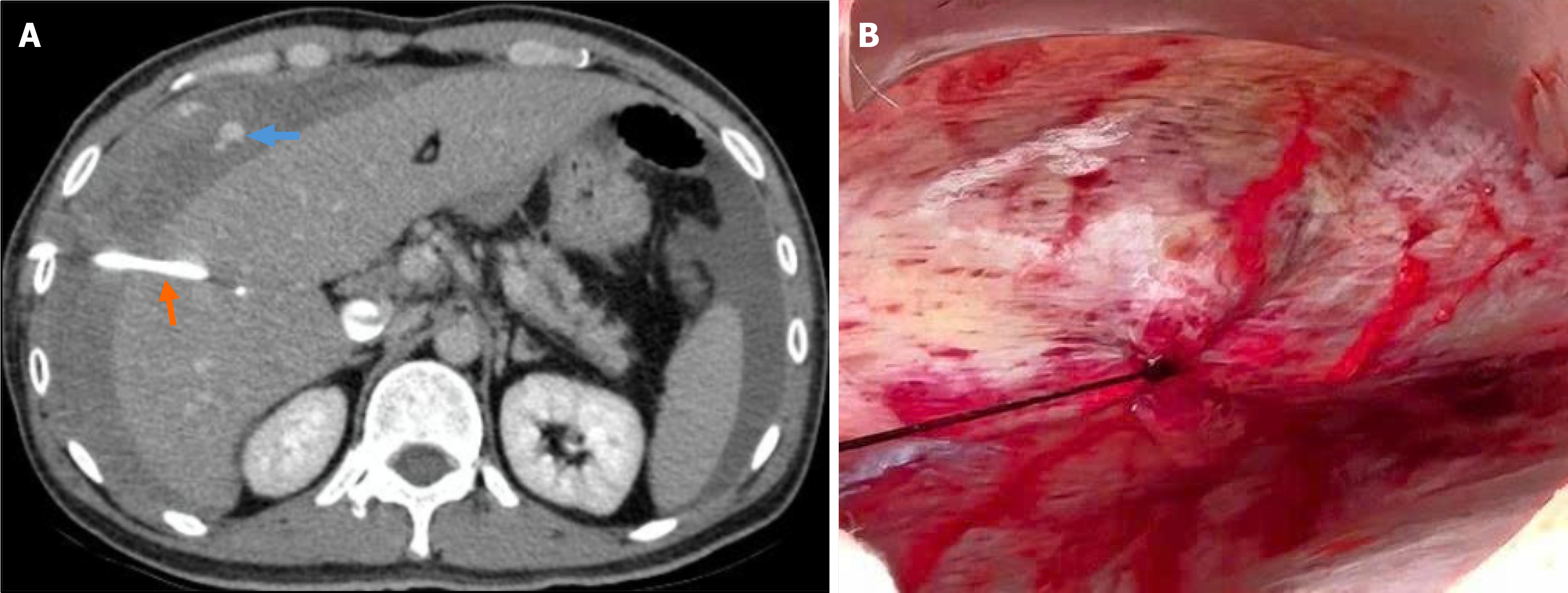
Figure 3 Extrahepatic intercostal hemorrhage after right intercostal percutaneous transhepatic biliary drainage.
A: Contrast-enhanced computed tomography demonstrates active extravasation along the access tract (blue arrow) adjacent to the percutaneous transhepatic biliary drainage catheter (orange arrow); B: Intraoperative image confirms chest-wall or intercostal bleeding.
- Citation: Le KL, Dinh TT, Thai KP, Pham TN, Tran MQ, Pham PC, Trinh MT, Duong NNQ, Nguyen TK, Le NK, Mai-Phan TA. Bleeding and vascular complications after percutaneous transhepatic biliary drainage: Incidence and associated factors in a Vietnamese tertiary center. World J Gastrointest Surg 2026; 18(7): 120929
- URL: https://www.wjgnet.com/1948-9366/full/v18/i7/120929.htm
- DOI: https://dx.doi.org/10.4240/wjgs.v18.i7.120929