
















Copyright: ©Author(s) 2026.
World J Gastrointest Surg. Jul 27, 2026; 18(7): 120207
Published online Jul 27, 2026. doi: 10.4240/wjgs.v18.i7.120207
Published online Jul 27, 2026. doi: 10.4240/wjgs.v18.i7.120207
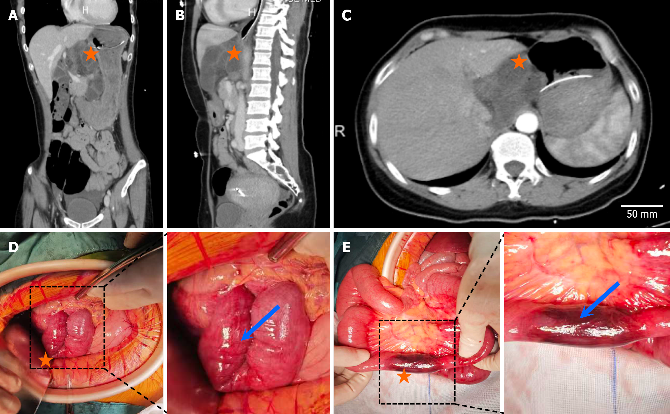
Figure 1 Multiplanar computed tomography images and intraoperative observations of internal hernia of case 1.
A-C: Multiplanar computed tomography images [coronal (A), sagittal (B), axial (C)] showing the location of the herniated lesion (orange star), consistent with the imaging features of internal hernia. The scale bar in (C) represents 50 mm; D and E: Intraoperative views: Gross appearance of the herniated bowel loops (indicated by the blue arrow) under laparoscopy; the dashed box denotes the target area, and the orange star marks the location of the herniated bowel (D). Intraoperative image after surgical manipulation (indicated by the blue arrow), showing the necrotic small intestine, with the orange star indicating the region of the necrotic bowel segment (E).
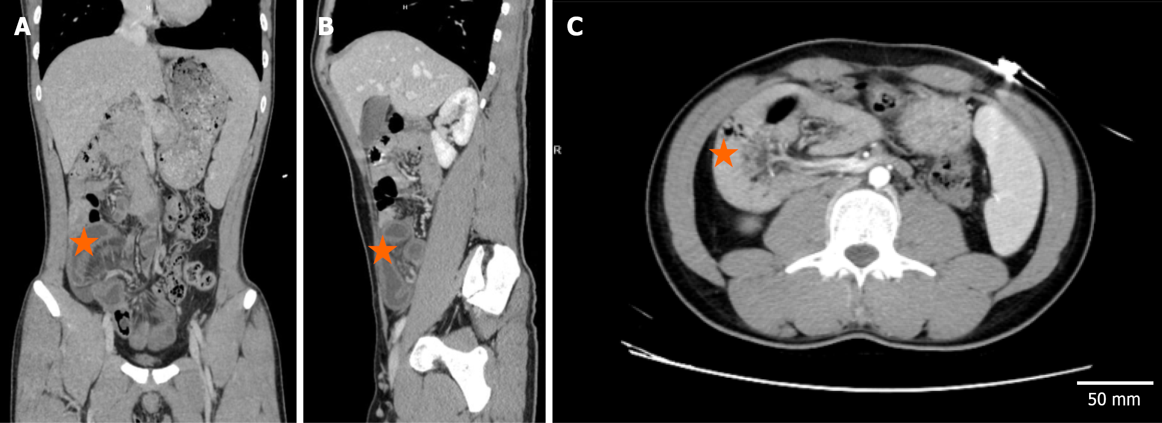
Figure 2 Multiplanar computed tomography images of case 2.
A: Coronal view; B: Sagittal view; C: Axial view. The area marked by the orange star indicates the herniated bowel loops, whose location and morphology are consistent with the imaging characteristics of internal hernia (bowel loops herniated into an abnormal space). The scale bar in (C) represents 50 mm.
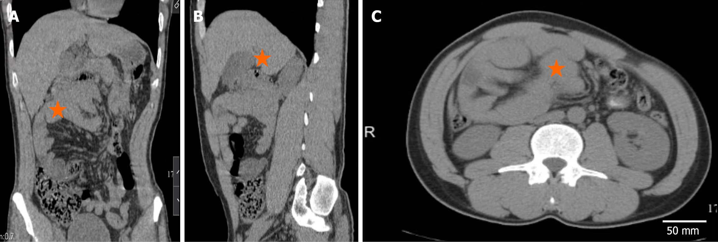
Figure 3 Multiplanar computed tomography images of case 3.
A: Coronal view; B: Sagittal view; C: Axial view. The area marked by the orange star indicates the herniated bowel loops, whose location and morphology are consistent with the imaging characteristics of internal hernia (bowel loops herniated into an abnormal space). The scale bar in (C) represents 50 mm.
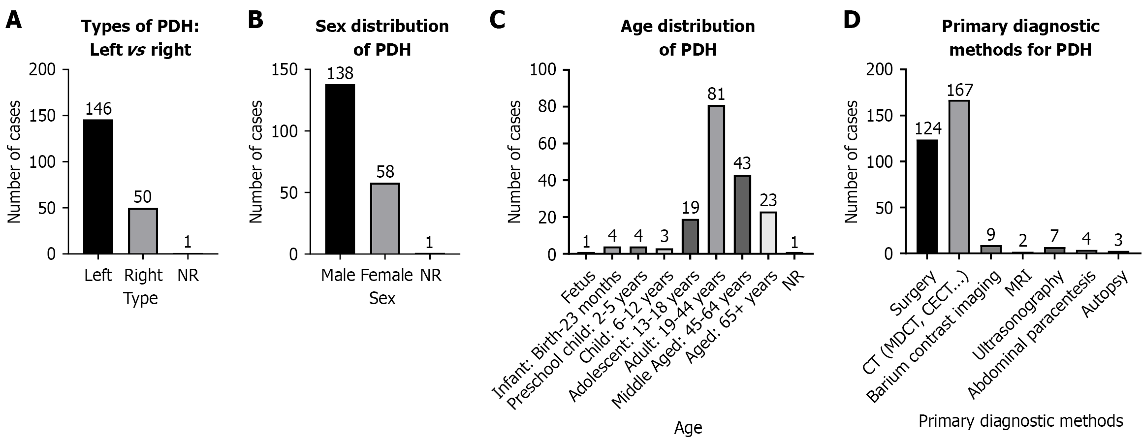
Figure 4 Epidemiological and clinical characteristics of paraduodenal hernia.
A: Paraduodenal hernia (PDH) showed a marked male predominance: 138 male cases vs 58 female cases, with 1 case of unclear gender, consistent with literature reports of male bias; B: Left-sided PDH was most common (n = 146), compared with 50 cases of right-sided PDH and 1 case of indeterminate laterality, reflecting anatomical predisposition to left-sided herniation; C: Highest incidence occurred in middle-aged adults (35-54 years, n = 81), followed by older adults (≥ 55 years, n = 43) and adolescents (13-18 years, n = 19). Cases were rare in infants and young children, suggesting a link to age-related abdominal changes or chronic intra-abdominal pressure; D: Cross-sectional imaging (computed tomo
- Citation: Chen LT, Pan BJ, Lu MD, Wang BH, Zhang N, Hu JW. Paraduodenal hernia with intestinal necrosis: Three case reports and review of literature. World J Gastrointest Surg 2026; 18(7): 120207
- URL: https://www.wjgnet.com/1948-9366/full/v18/i7/120207.htm
- DOI: https://dx.doi.org/10.4240/wjgs.v18.i7.120207