
















Copyright: ©Author(s) 2026.
World J Gastrointest Surg. Jul 27, 2026; 18(7): 120053
Published online Jul 27, 2026. doi: 10.4240/wjgs.120053
Published online Jul 27, 2026. doi: 10.4240/wjgs.120053
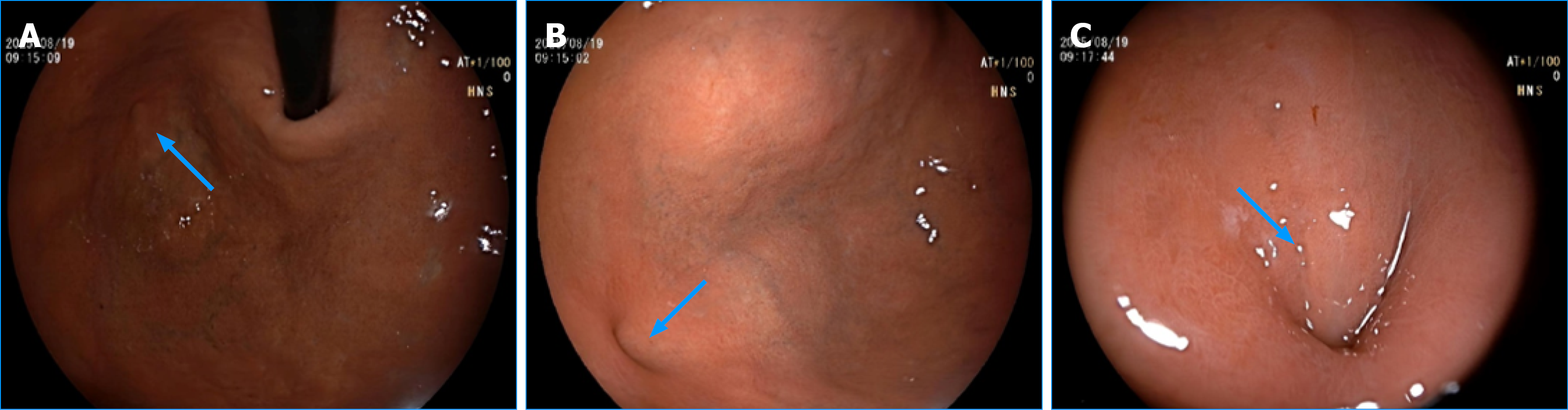
Figure 1 Endoscopic view of the fundic orifice of a gastrojejunal fistula (arrows).
A small, smooth orifice on the posterior fundus with no surrounding ulceration. A-C: The same lesion viewed from different distances and angles under endoscopy.
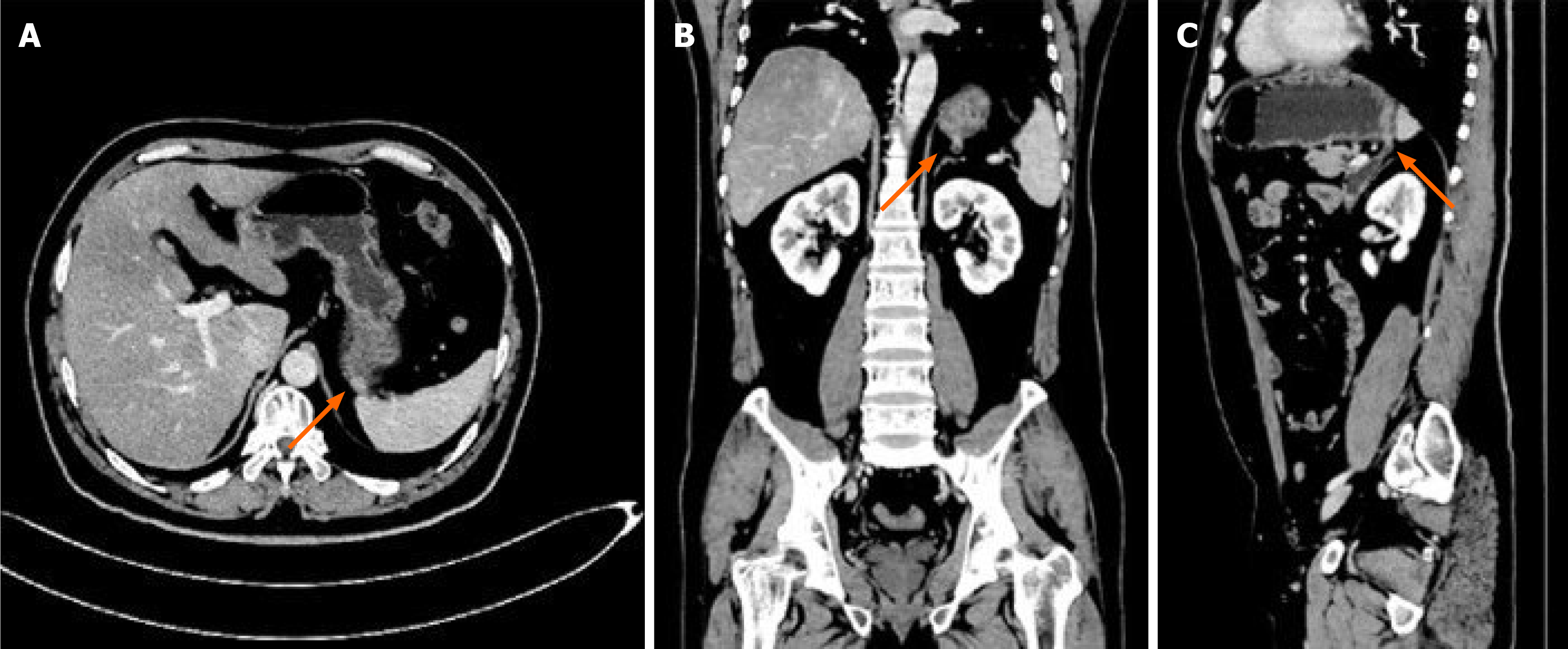
Figure 2 Contrast-enhanced computed tomography showing the fistula (arrows).
A: Axial view; B: Coronal view; C: Sagittal view. A focal defect in the posterior fundic wall communicates with an adjacent jejunal loop; fat planes are preserved, without inflammatory stranding.
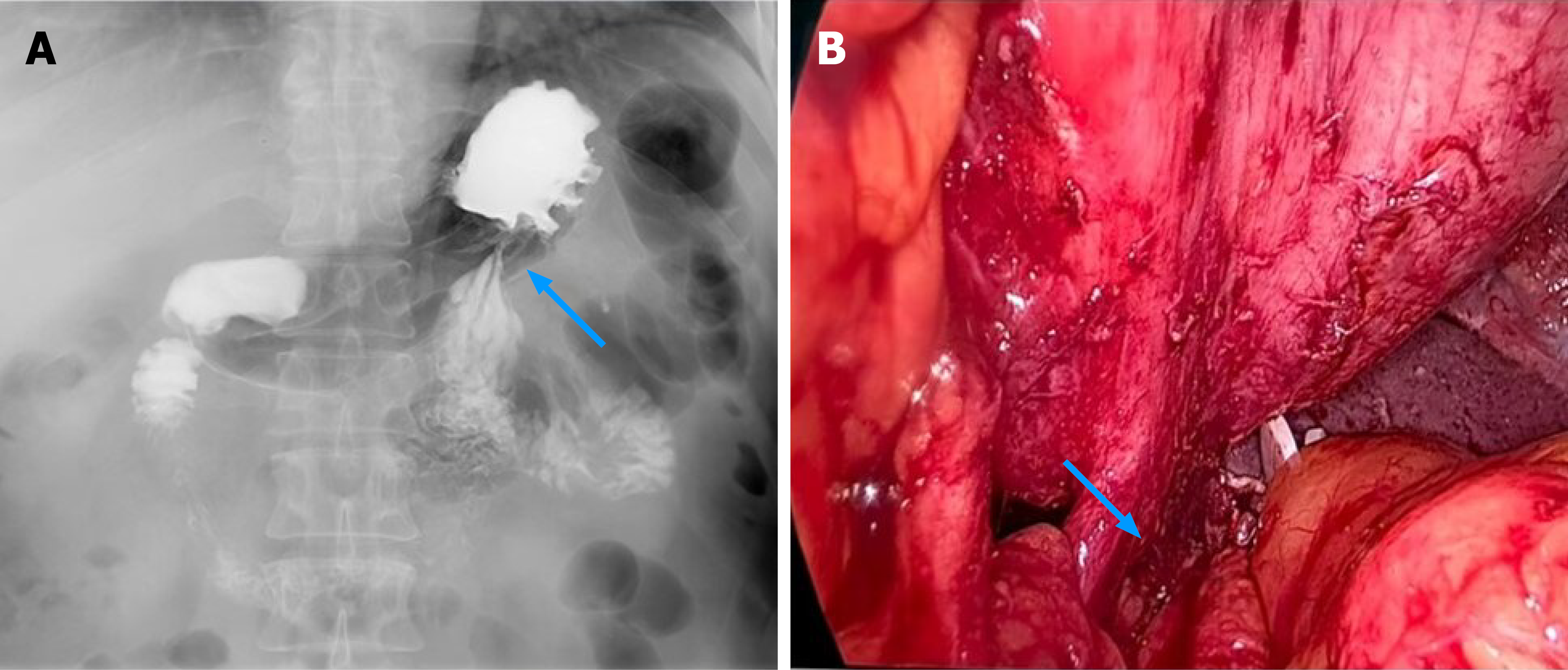
Figure 3 Appearance of the fistulous tract on upper gastrointestinal contrast study and diagnostic laparoscopy (arrows).
A: Upper gastrointestinal contrast study. Jejunum opacifies from the stomach, confirming the communication; B: Diagnostic laparoscopy. A smooth, isolated, cord-like tract arises approximately 5 cm distal to the ligament of Treitz, courses through the transverse mesocolon and behind the pancreas to the fundus, with no adhesions.
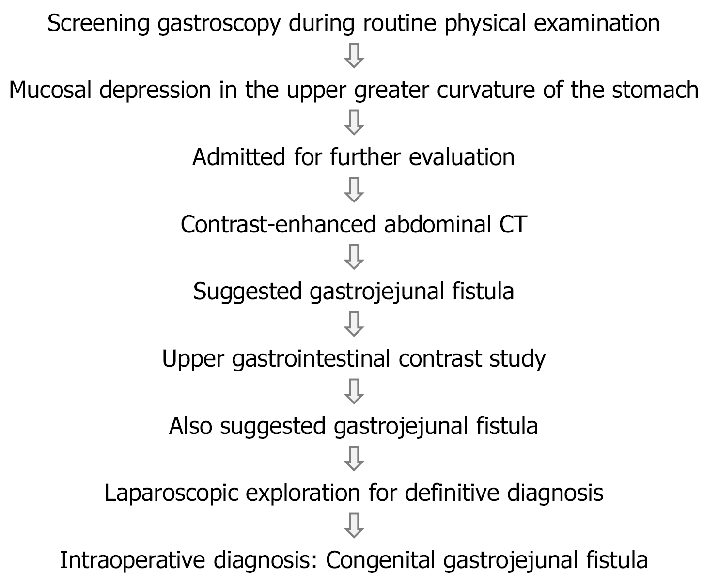
Figure 4 Diagnostic workflow.
A 56-year-old asymptomatic male underwent screening gastroscopy during a routine physical examination, which revealed a mucosal depression in the upper greater curvature of the stomach. Subsequent imaging and laparoscopy confirmed a congenital gastrojejunal fistula. CT: Computed tomography.
- Citation: Chen XR, Guo HY, Liang R, Zhang CY, Yuan XB, Wang XF. Congenital gastrojejunal fistula in an asymptomatic adult: A case report. World J Gastrointest Surg 2026; 18(7): 120053
- URL: https://www.wjgnet.com/1948-9366/full/v18/i7/120053.htm
- DOI: https://dx.doi.org/10.4240/wjgs.120053