
















Copyright: ©Author(s) 2026.
World J Gastrointest Surg. Jul 27, 2026; 18(7): 119297
Published online Jul 27, 2026. doi: 10.4240/wjgs.v18.i7.119297
Published online Jul 27, 2026. doi: 10.4240/wjgs.v18.i7.119297
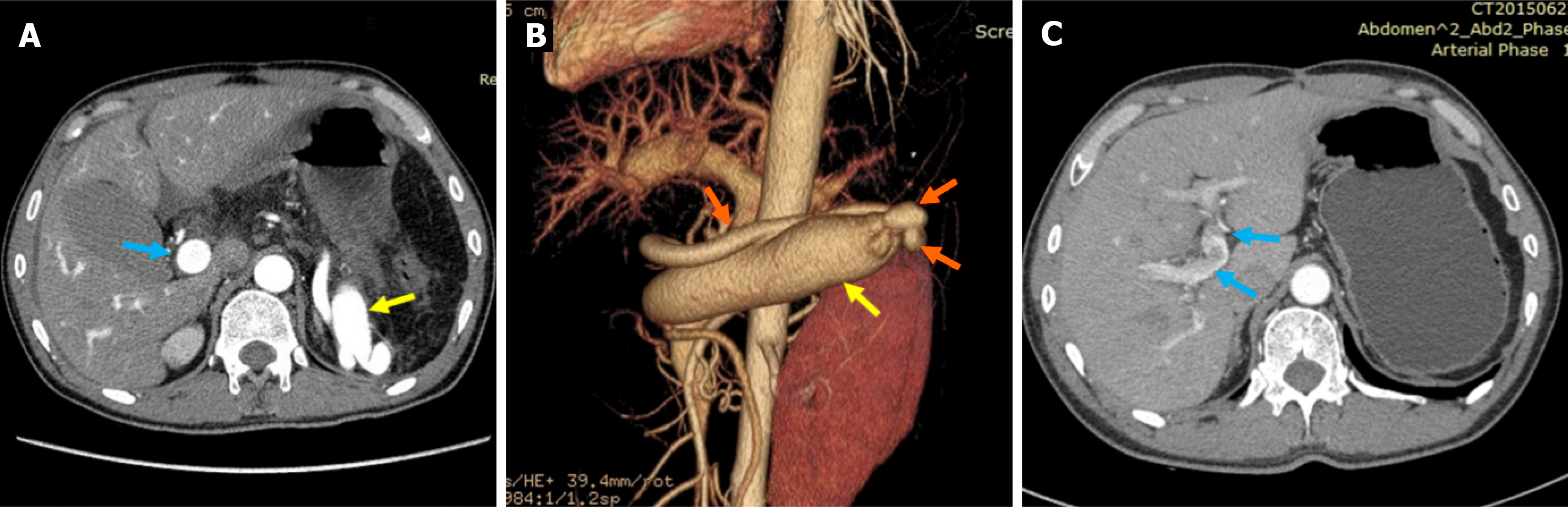
Figure 1 Abdominal computed tomography angiography findings in splenic arteriovenous fistula.
A: Abdominal computed tomography (CT) angiography shows dilation and early filling of splenic vein (yellow arrow) and portal vein (blue arrow) in arterial phase; B: Three-dimensional reconstruction CT showed a fistula between the splenic artery (orange arrow) and the dilated splenic vein (yellow arrow); C: Abdominal CT angiography shows no early filling of portal vein (blue arrow) in arterial phase 8 months later.
- Citation: Yan JY, Wang JY, Chen W, Hua R. Acute portal hypertension due to delayed post-splenectomy arteriovenous fistula: A case report. World J Gastrointest Surg 2026; 18(7): 119297
- URL: https://www.wjgnet.com/1948-9366/full/v18/i7/119297.htm
- DOI: https://dx.doi.org/10.4240/wjgs.v18.i7.119297