
















Copyright: ©Author(s) 2026.
World J Gastrointest Surg. May 27, 2026; 18(5): 119504
Published online May 27, 2026. doi: 10.4240/wjgs.v18.i5.119504
Published online May 27, 2026. doi: 10.4240/wjgs.v18.i5.119504
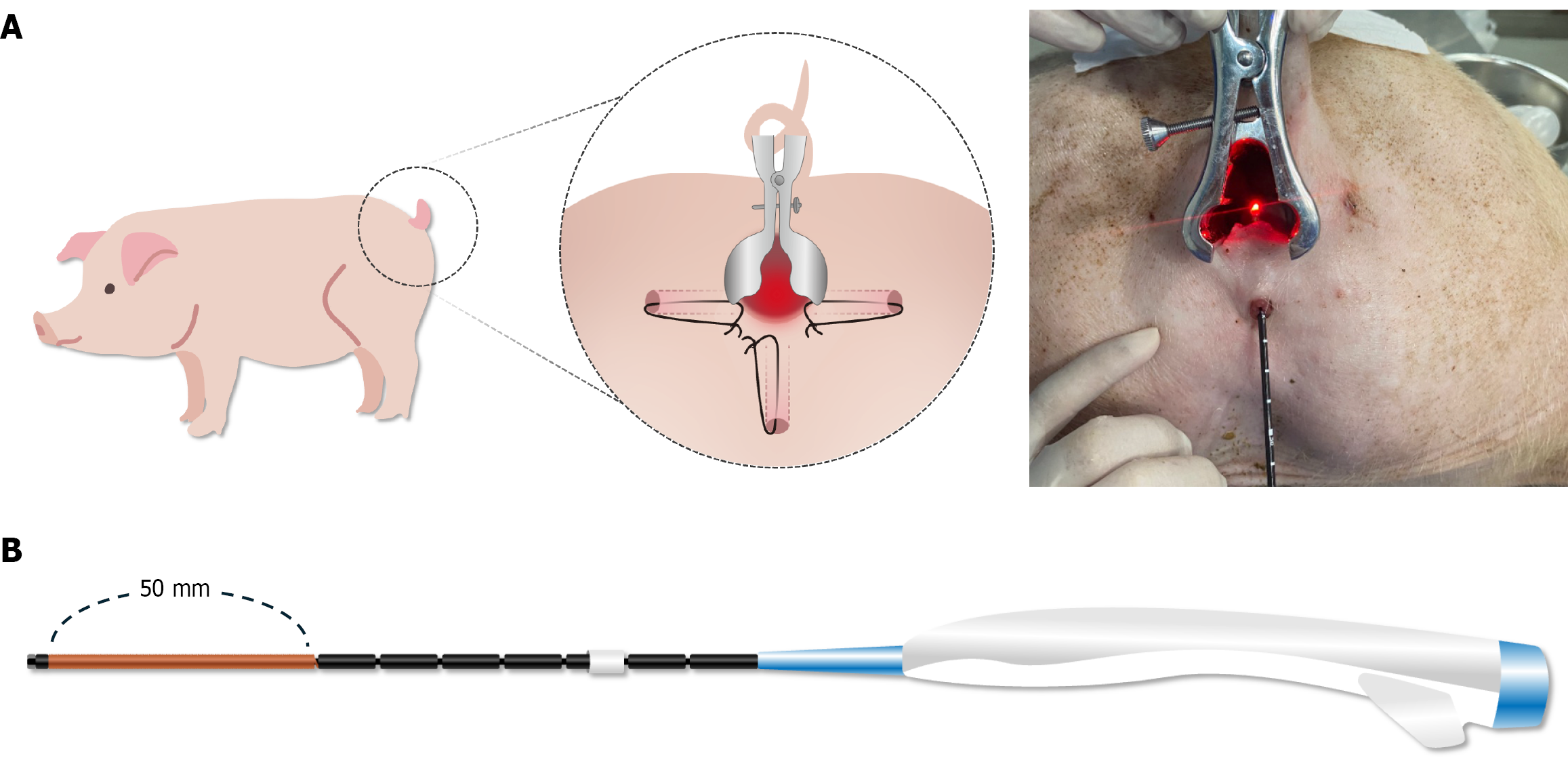
Figure 1 Experimental set-up and novel radiofrequency ablation device.
A: Schematic of the porcine fistula model used for in-vivo optimization of radiofrequency ablation (RFA) parameters. Three fistula tracts were surgically created in each animal and kept patent with setons, as illustrated in the enlarged inset. For ablation, seton was removed and the RFA catheter introduced endoluminally, as confirmed by the intra-operative photograph; B: Prototype segmental RFA catheter. The distal 50 mm copper-colored segment constitutes the active ablation blade, and graduations at 10 mm intervals along the shaft allow precise depth control. This 50 mm active segment delivers a uniformly distributed ablation along its entire length, thereby overcoming the focal-tip limitation of conventional devices.
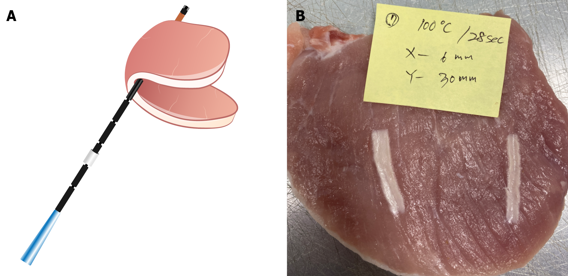
Figure 2 Ex vivo platform for preliminary screening radiofrequency ablation temperature-time settings.
A: Schematic of the segmental radiofrequency ablation catheter positioned along the midline of a fresh porcine loin; the loin was then folded over the catheter so that the loin enveloped the electrode and maintained continuous, uniform contact during energy delivery; B: Loin re-opened after ablation (representative condition: 100 °C/28 seconds), showing a well-demarcated linear ablation tract. This configuration enabled direct caliper measurement of ablation depth and length for each candidate settings.
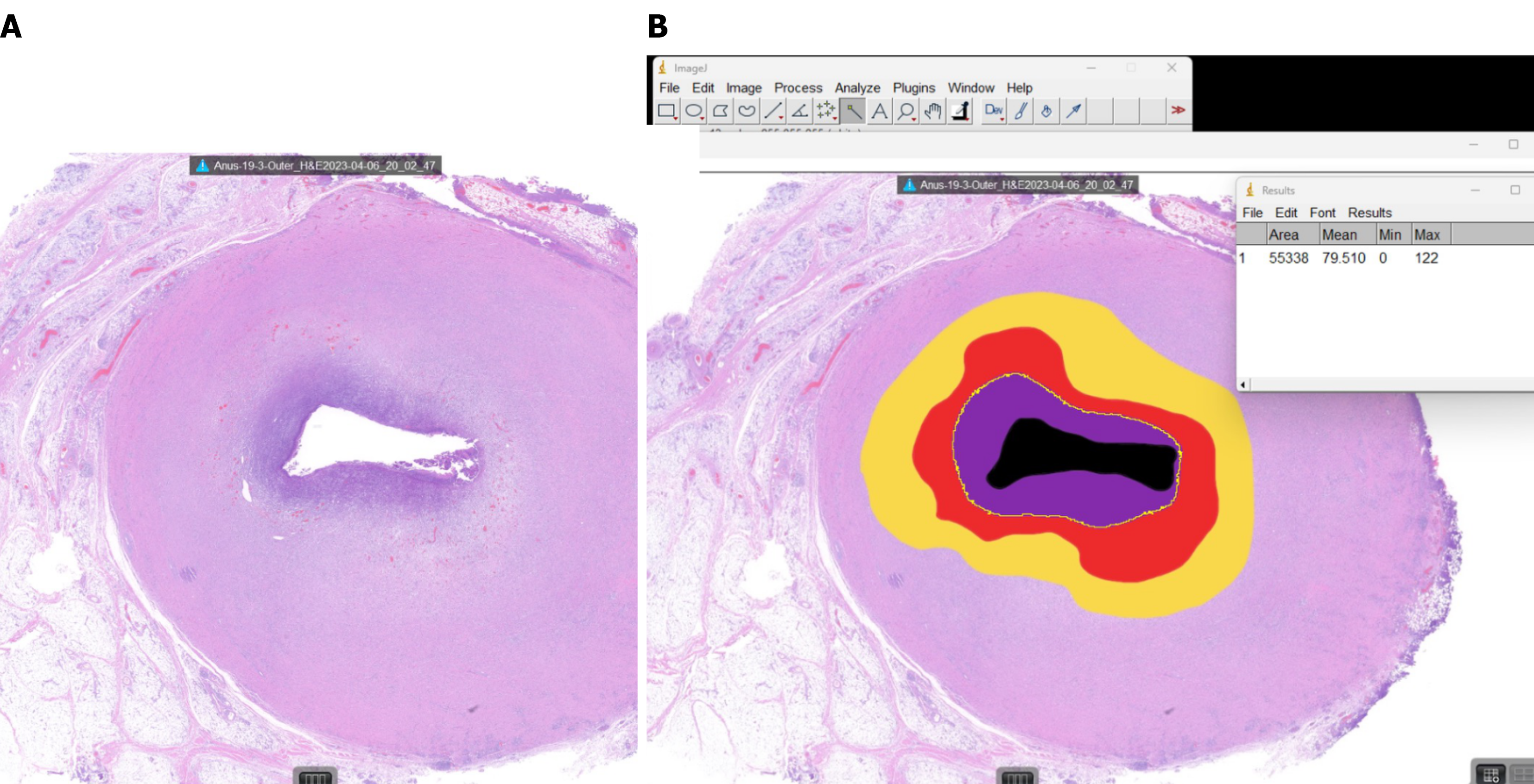
Figure 3 Representative histological section illustrating Image J-based quantification of ablation and granulation tissue.
A: Hematoxylin and eosin -stained cross-section of an outer fistula-tract segment harvested after radiofrequency ablation ablation (120 °C/24 seconds), showing a centrally obliterated lumen surrounded by a concentric zone of thermal injury; B: The same section processed in Image J, with color overlays delineating tissue compartments used for morphometry: Residual lumen (black), carbonized/charred core (violet), coagulative necrosis (red), transition zone (orange) and residual granulation tissue (yellow).
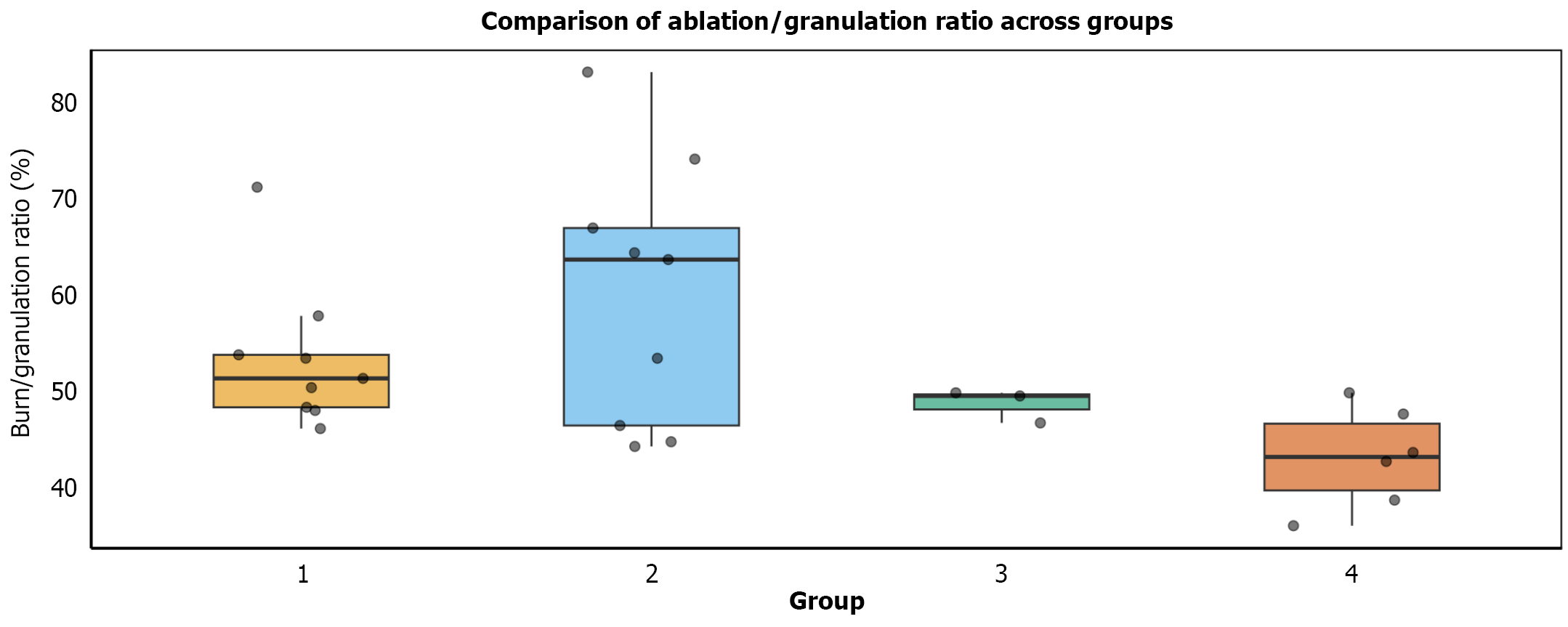
Figure 4 Box plot illustrating the ablation-to-granulation tissue area ratios across the four experimental groups.
Group 2 (120 °C for 24 seconds) exhibited the highest ablation/granulation ratios. Group 2 was significantly higher than groups 1, 3, and 4 by Tukey’s honestly significant difference post hoc test (all P < 0.001).
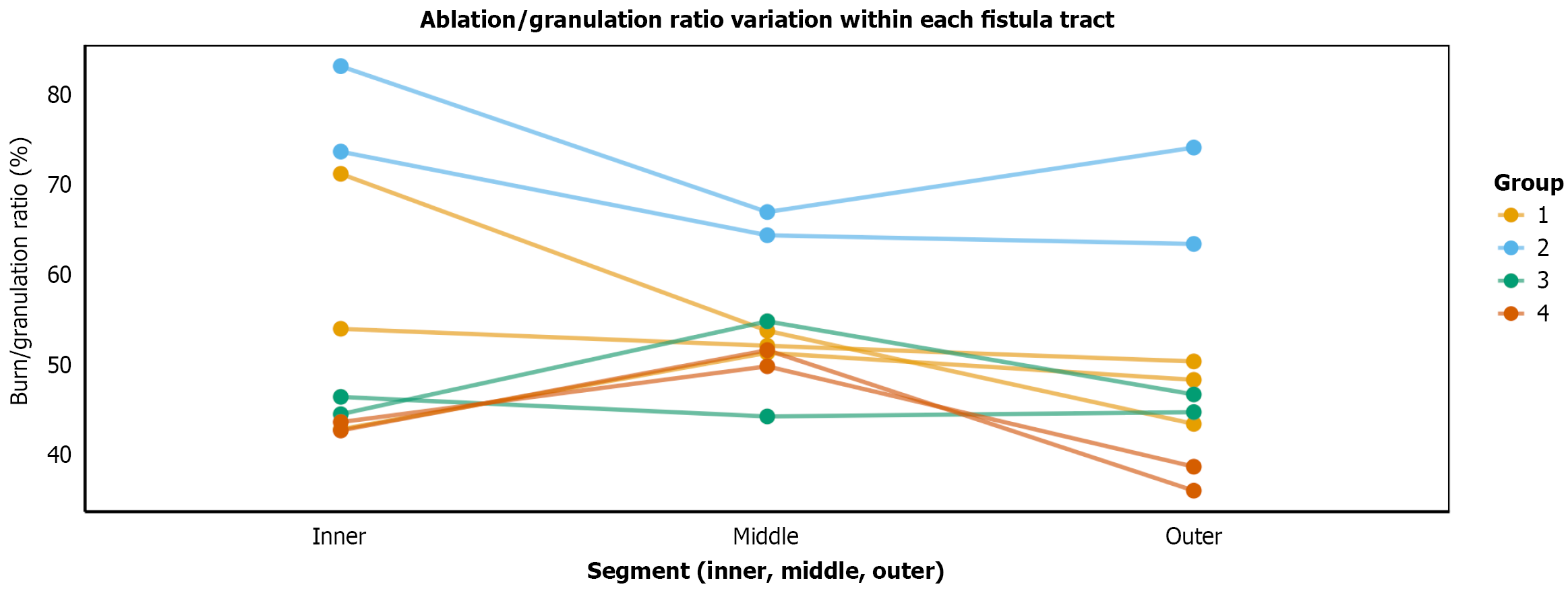
Figure 5 Line plot depicting the ablation-to-granulation tissue ratios within individual fistula tracts, segmented into inner, middle, and outer regions.
No consistent pattern of variation was observed, further supporting the uniformity of the radiofrequency ablation effect across the tract.
- Citation: Yoon S, Ji WB, Um JW, Hong KD. Optimizing radiofrequency ablation parameters for perianal fistula treatment: A porcine model study. World J Gastrointest Surg 2026; 18(5): 119504
- URL: https://www.wjgnet.com/1948-9366/full/v18/i5/119504.htm
- DOI: https://dx.doi.org/10.4240/wjgs.v18.i5.119504