
















Copyright: ©Author(s) 2026.
World J Gastrointest Surg. May 27, 2026; 18(5): 119094
Published online May 27, 2026. doi: 10.4240/wjgs.v18.i5.119094
Published online May 27, 2026. doi: 10.4240/wjgs.v18.i5.119094
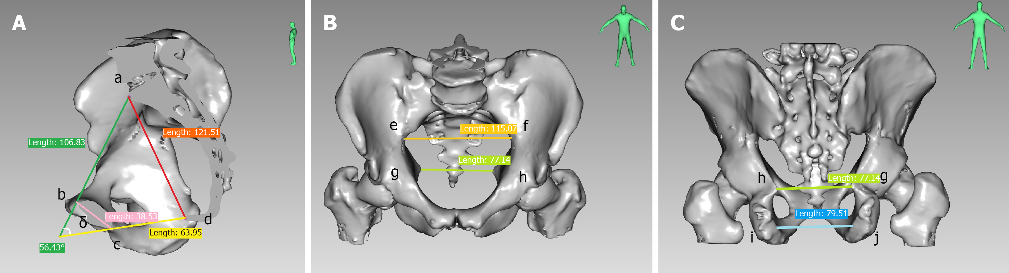
Figure 1 Pelvic measurements for three-dimensional reconstruction.
A: Anterior-posterior diameter of pelvic inlet (ab); anterior-posterior diameter of pelvic outlet (cd); superior-inferior diameter of the pubic symphysis (bc); sacrococcygeal distance (ad); and sacrococcygeal-pubic angle (δ); B: Transverse diameter of pelvic inlet (ef) and interischial spine diameter (gh); C: Interischial spine diameter (gh) and interischial tuberosity diameter (ij).
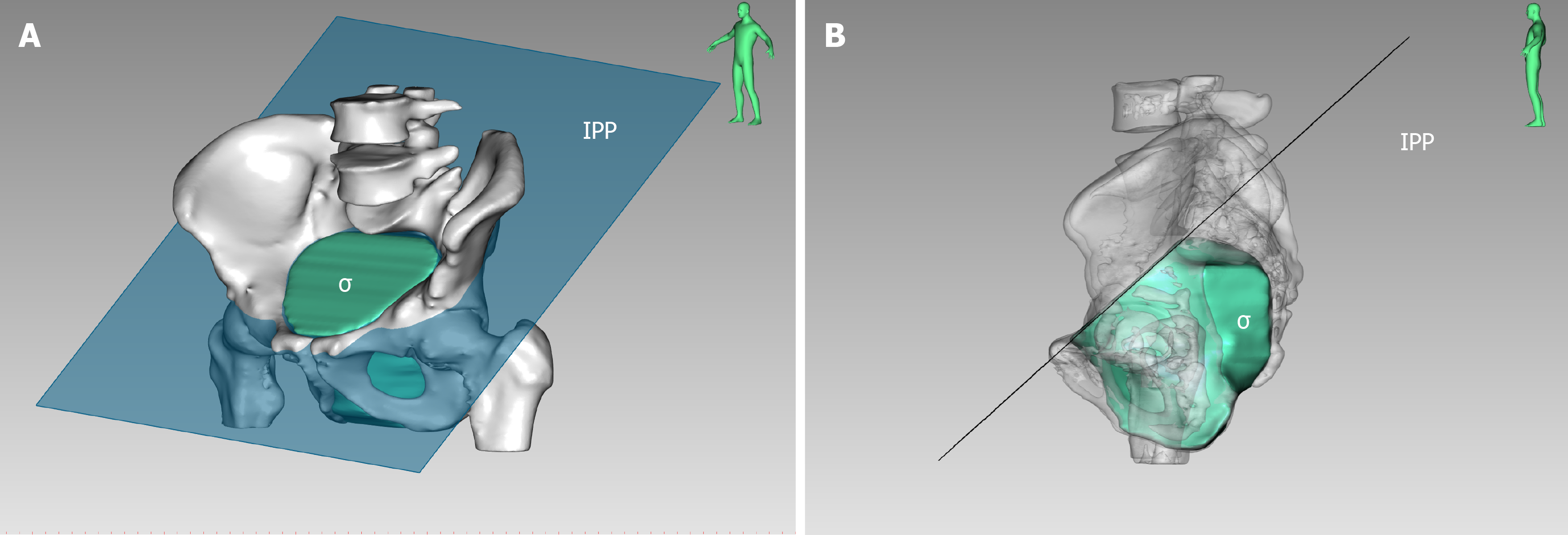
Figure 2 Three-dimensional reconstruction of the pelvis: Pelvic volume and inlet pelvic plane.
A: Anterolateral view: Pelvic volume (σ) and inlet pelvic plane (IPP); B: Lateral view: Pelvic volume (σ) and IPP (semi-translucent pelvis). IPP: Inlet pelvic plane.
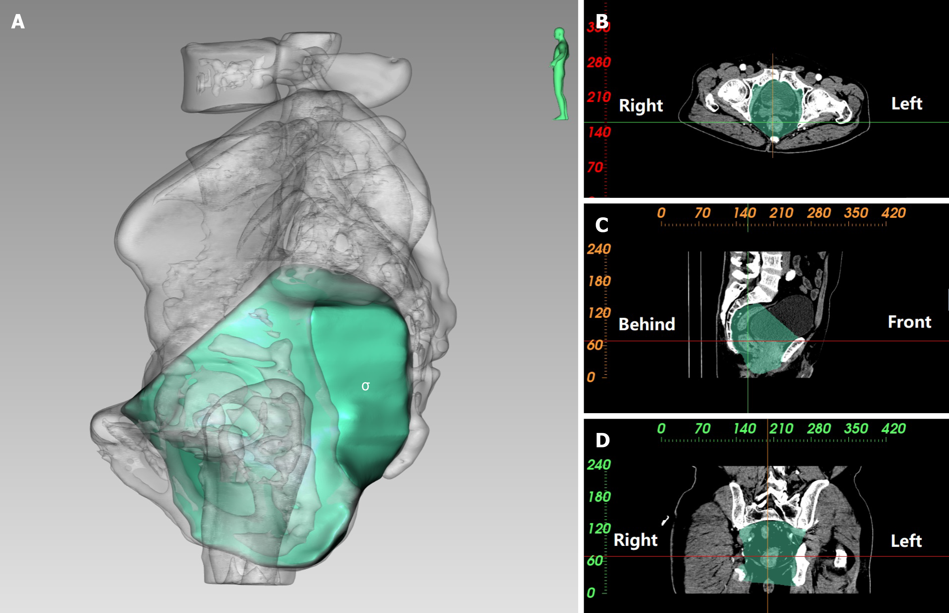
Figure 3 Three-dimensional reconstruction of pelvic volume.
A: Lateral view: Pelvic volume (σ; semi-translucent pelvis); B: Pelvic volume on the transverse plane; C: Pelvic volume on the sagittal plane; D: Pelvic volume on the coronal plane.
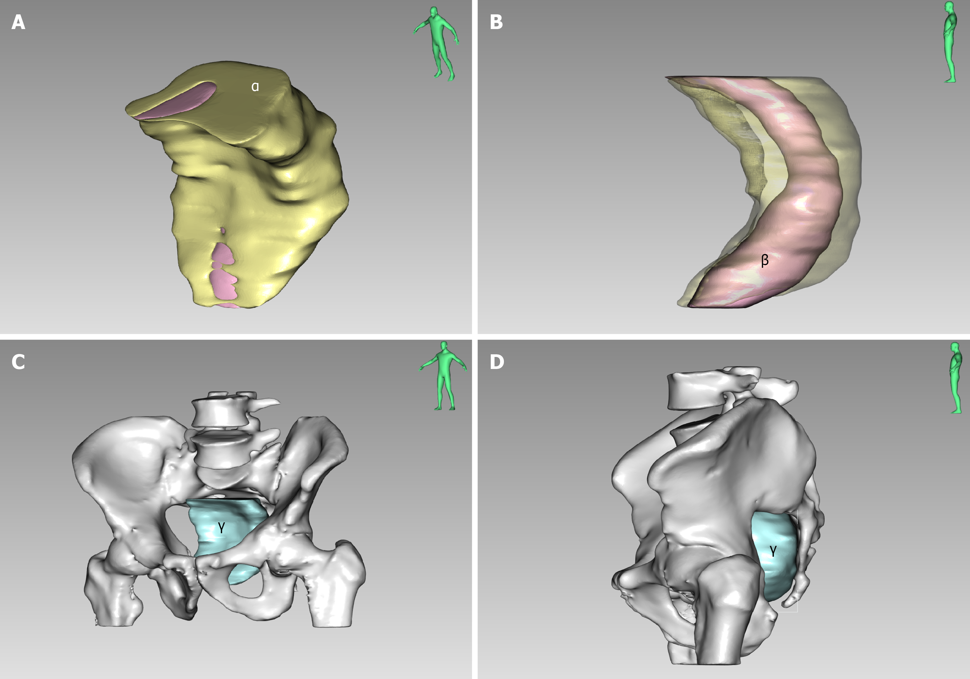
Figure 4 Three-dimensional reconstruction of rectal mesenteric-related volumes in different orientations.
A: Anterolateral view: Rectal mesenteric fat at the level of the superior border of the third sacral vertebra (α); B: Lateral view: Rectal volume at the level of the superior border of the third sacral vertebra (β; semi-translucent rectal mesenteric fat); C: Anterolateral view: Rectal mesenteric volume at the level of the superior border of the third sacral vertebra ((γ = α + β) and pelvis; D: Lateral view: Rectal mesenteric volume at the level of the superior border of the third sacral vertebra (γ) and pelvis.
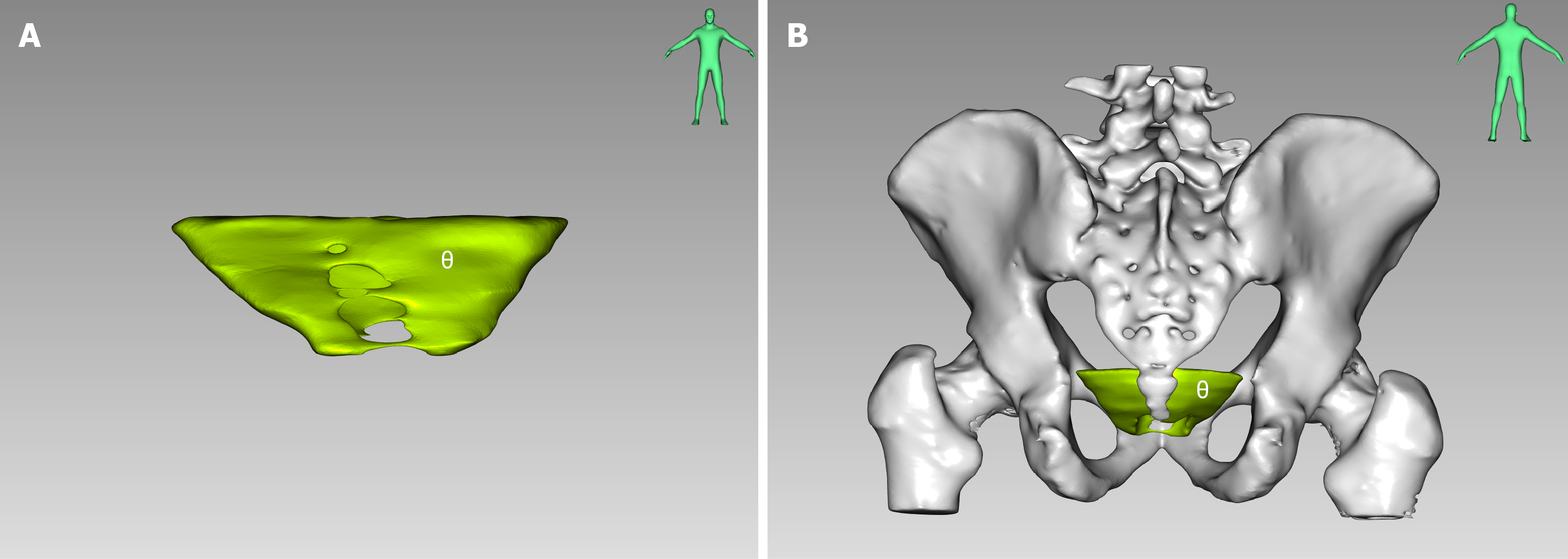
Figure 5 Three-dimensional reconstruction of rectal mesenteric fat at the level of the sciatic spine.
A: Front view: Rectal mesenteric fat at the level of the sciatic spine (θ); B: Behind view: Rectal mesenteric fat at the level of the sciatic spine (θ) and the pelvis.
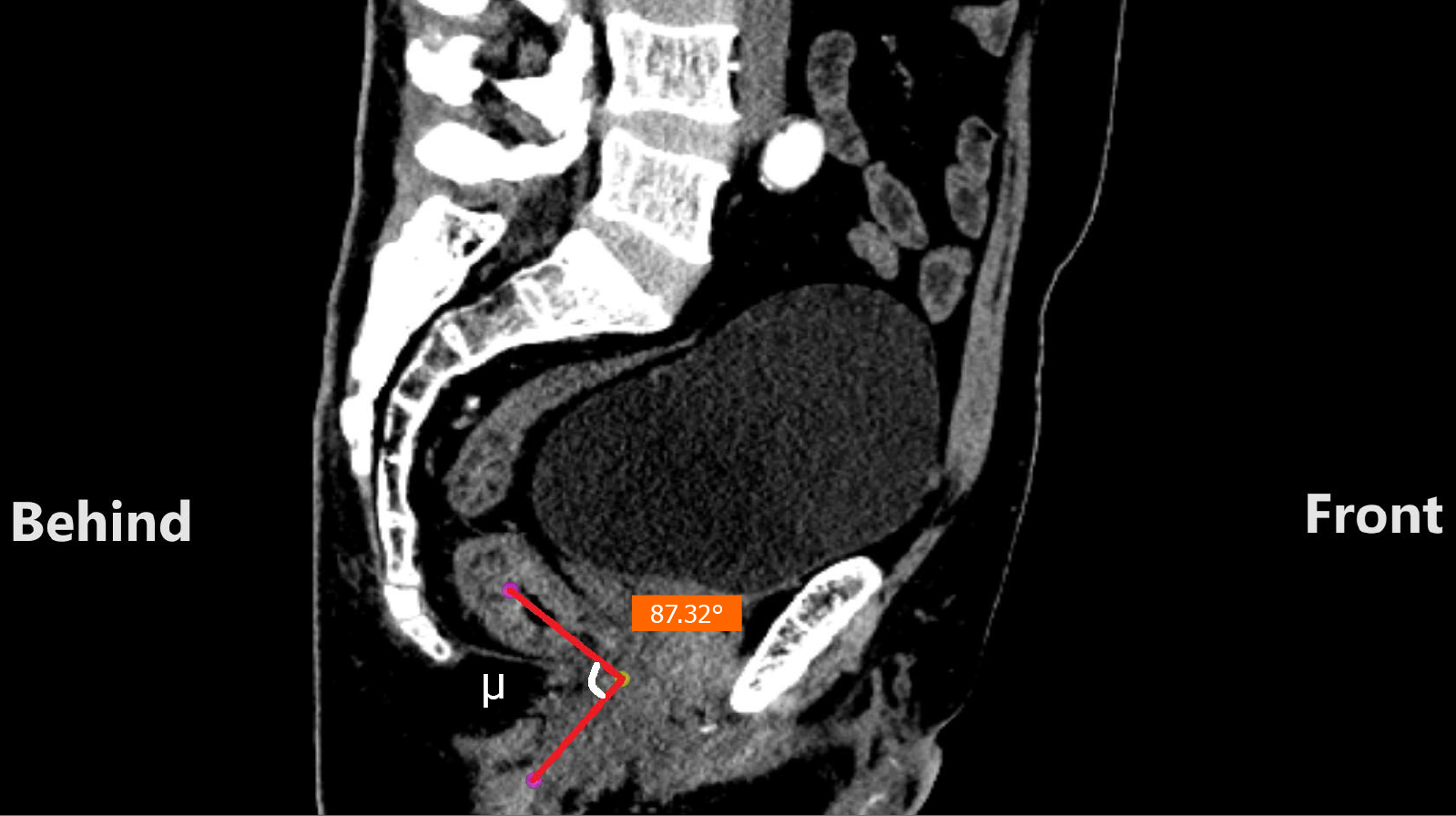
Figure 6 Anorectal angle on the median sagittal plane.
Anorectal angle (μ).
- Citation: Ke FY, Chen H, Liu JS, Li WC, Dhamija G, Viroja RD, Chen GP, Zhou XC. Clinical value of computed tomography-based three-dimensional reconstruction of the pelvis and mesorectum in middle to low rectal carcinoma. World J Gastrointest Surg 2026; 18(5): 119094
- URL: https://www.wjgnet.com/1948-9366/full/v18/i5/119094.htm
- DOI: https://dx.doi.org/10.4240/wjgs.v18.i5.119094