
















Copyright: ©Author(s) 2026.
World J Gastrointest Surg. May 27, 2026; 18(5): 118166
Published online May 27, 2026. doi: 10.4240/wjgs.v18.i5.118166
Published online May 27, 2026. doi: 10.4240/wjgs.v18.i5.118166
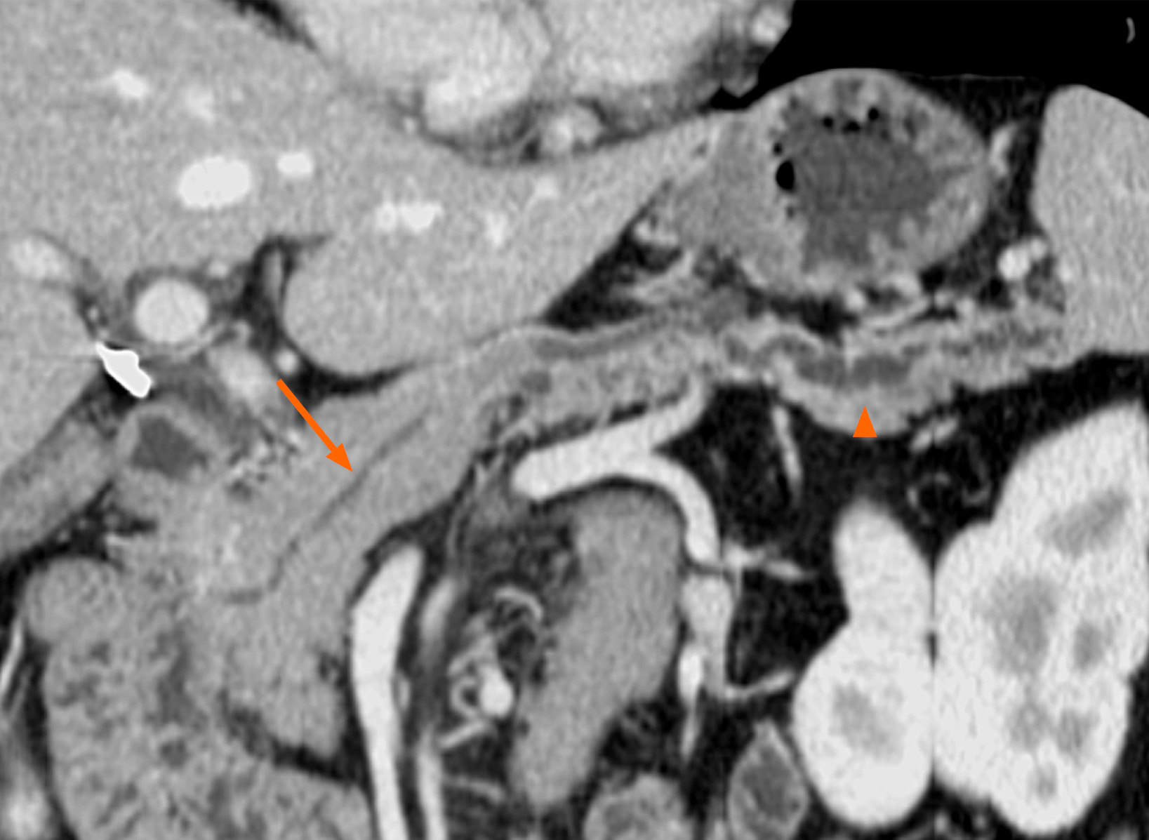
Figure 1 Preoperative imaging before the initial surgery.
Contrast-enhanced computed tomography image obtained in March 2005, before the initial surgery. The main pancreatic duct in the body and tail of the pancreas is dilated (orange arrowhead), whereas no dilatation is observed in the pancreatic head (orange arrow). These findings led to the diagnosis of main duct-type intraductal papillary mucinous neoplasm in the pancreatic body and tail.
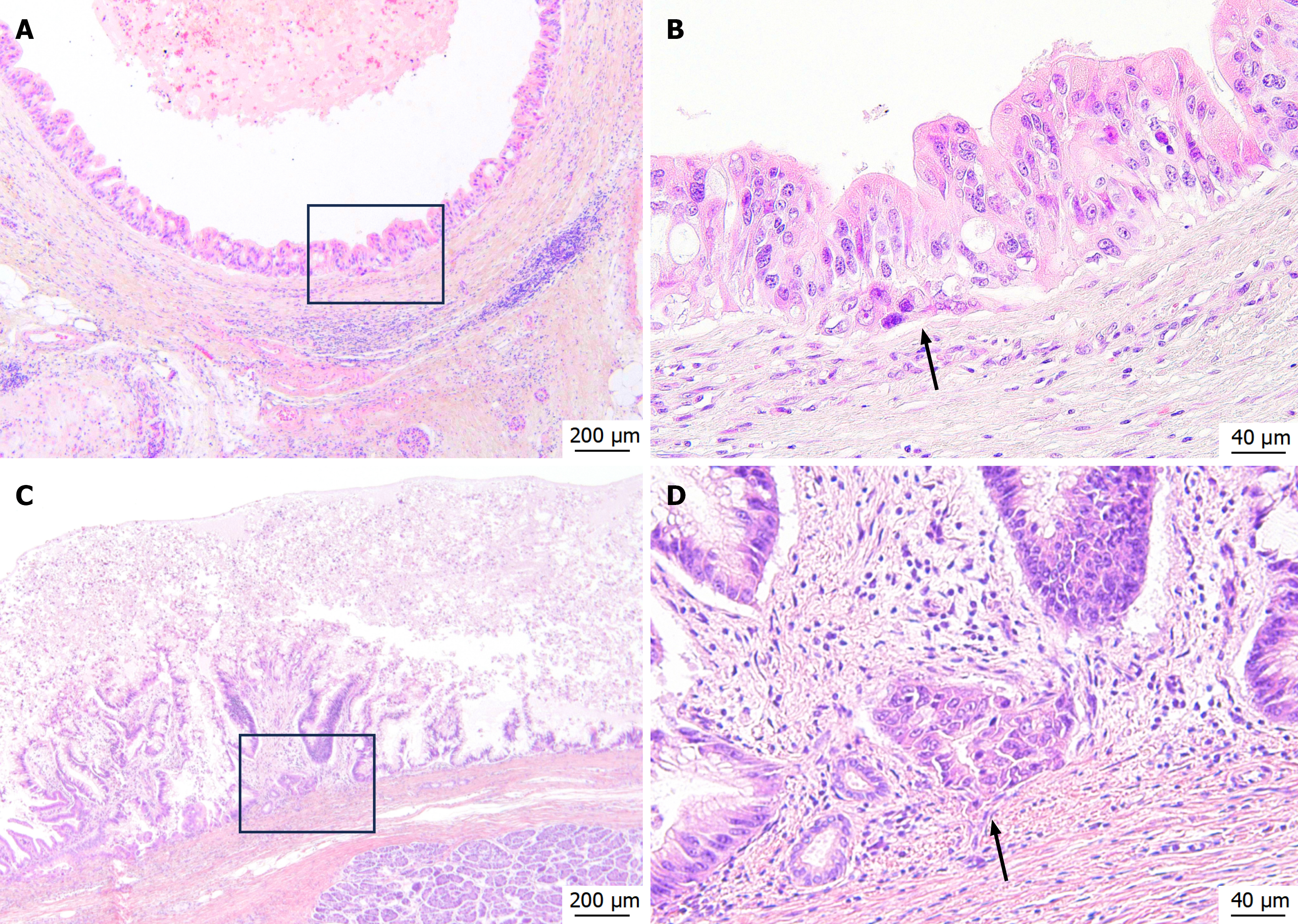
Figure 2 Histopathological findings of microinvasive intraductal papillary mucinous carcinoma (Hematoxylin and eosin staining).
A and B: Histopathological images from the first surgery. Atypical columnar epithelium showing dysplasia proliferates in papillary patterns within the dilated pancreatic duct. Slight irregularity of the basal aspect indicating microinvasion is observed in the marginal area (black arrow) (A: × 40; B: × 200); C and D: Histopathological images from the second surgery. Findings similar to those of the first surgery, including microinvasion (black arrow), are observed (C: × 40; D: × 200).
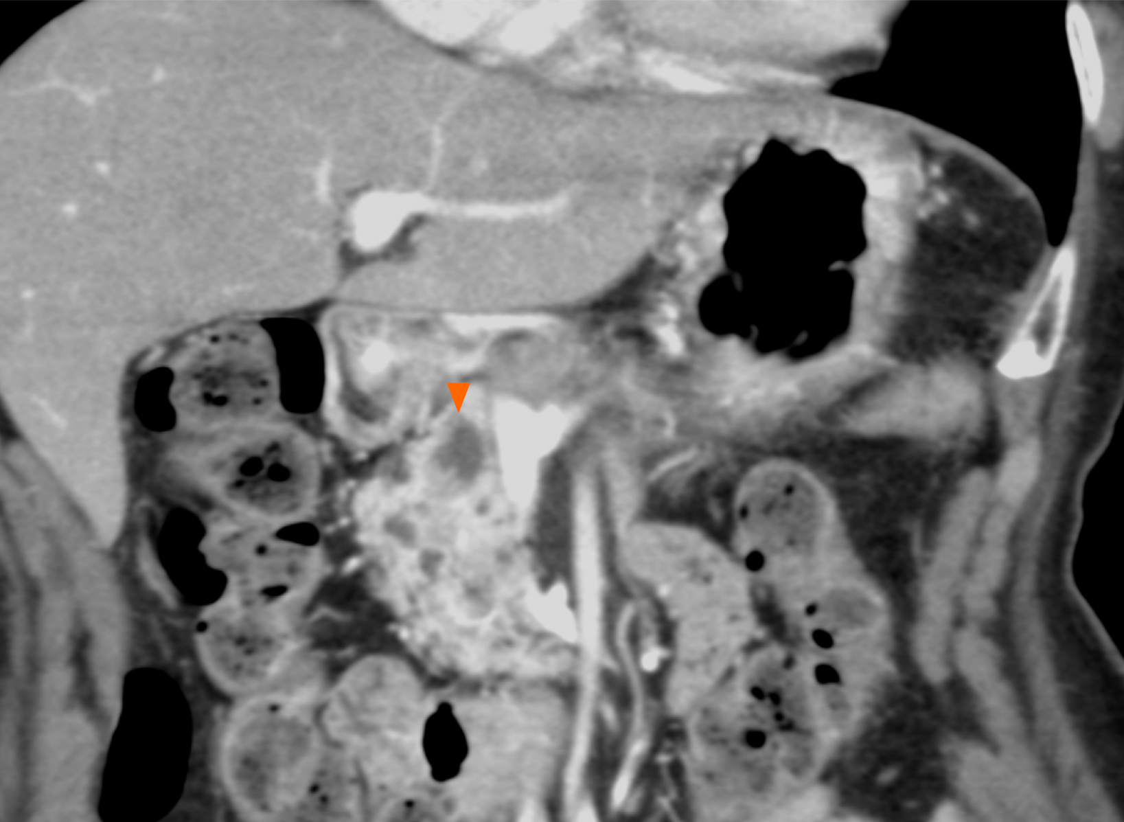
Figure 3 Imaging findings before the second surgery.
Contrast-enhanced computed tomography image obtained in June 2007 before the second surgery. The main pancreatic duct in the remnant pancreatic head is dilated (orange arrowhead), leading to the diagnosis of main duct-type intraductal papillary mucinous neoplasm in the remnant pancreatic head.
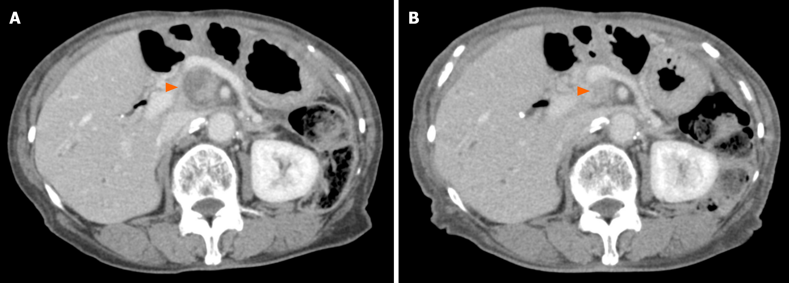
Figure 4 Radiologic detection and chemotherapy response of lymph node recurrence.
A: Contrast-enhanced computed tomography (CE-CT) in January 2020 shows a 27 mm × 22 mm mixed solid-mucinous mass right of the superior mesenteric artery (orange arrowhead), with elevated carbohydrate antigen 19-9 and duodenal pancreatic cancer antigen-2 indicating lymph node recurrence of microinvasive intraductal papillary mucinous carcinoma; B: CE-CT in March 2022 demonstrates complete resolution of the lymph node recurrence (orange arrowhead) and normalization of tumor markers, consistent with a favorable response to S-1 therapy.
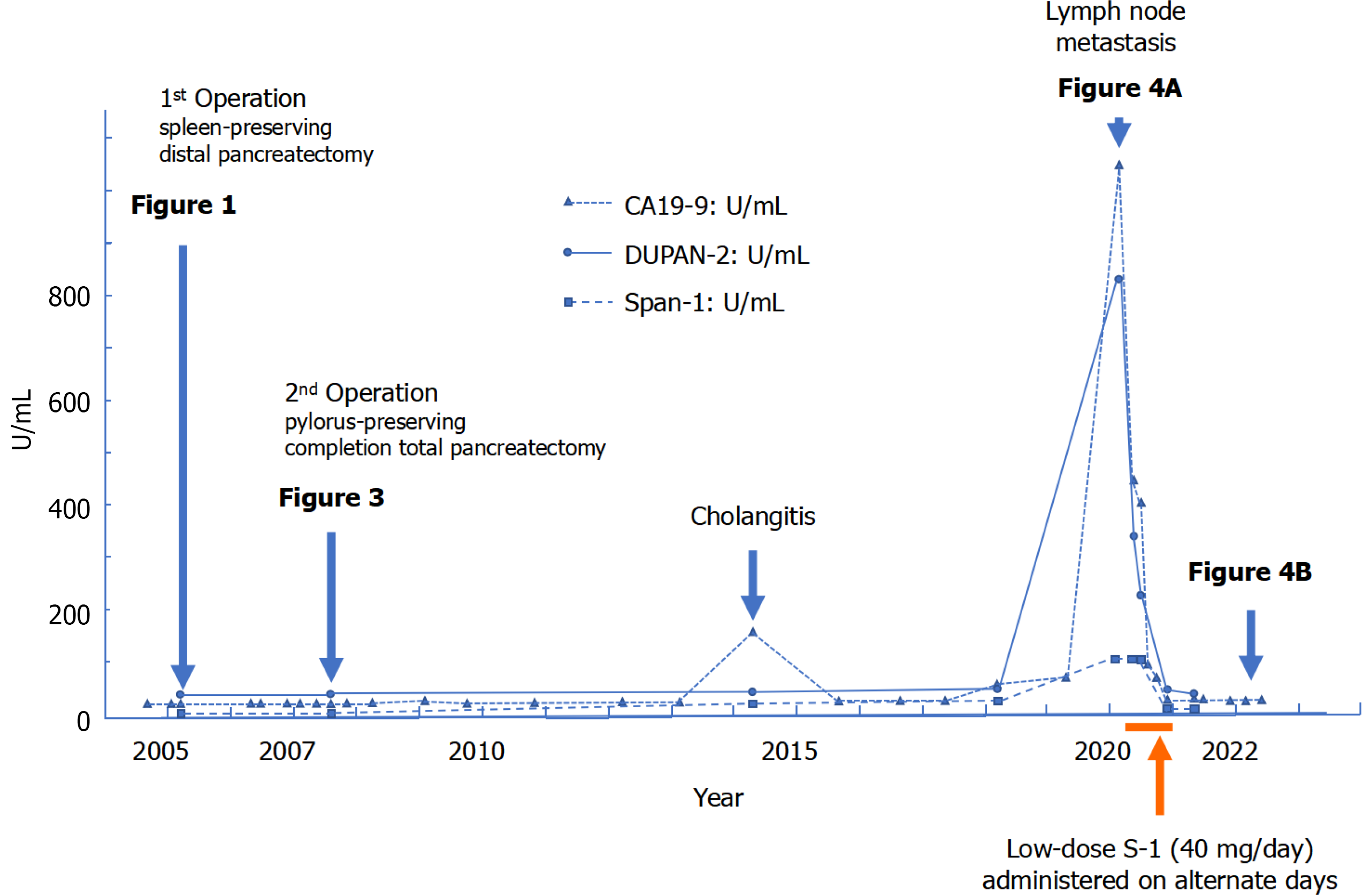
Figure 5 Clinical course and tumor marker trends.
The patient’s clinical course is described below. Serum tumor marker levels (carbohydrate antigen 19-9, duodenal pancreatic cancer antigen-2, and S-pancreas antigen-1) are plotted as line graphs. Major clinical events, including surgery and hospitalization for cholangitis, are indicated. Thirteen years after the second surgery, marked elevation of tumor markers was observed at the time of lymph node recurrence adjacent to the superior mesenteric artery. Following 9 months of alternate-day oral administration of S-1, all tumor marker levels returned to within normal ranges. CA19-9: Carbohydrate antigen 19-9; DUPAN-2: Duodenal pancreatic cancer antigen-2; Span-1: S-pancreas antigen-1.
- Citation: Nomoto K, Hishida M, Hayashi M, Nomoto S. Late nodal recurrence after two curative resections for microinvasive intraductal papillary mucinous carcinoma: A case report. World J Gastrointest Surg 2026; 18(5): 118166
- URL: https://www.wjgnet.com/1948-9366/full/v18/i5/118166.htm
- DOI: https://dx.doi.org/10.4240/wjgs.v18.i5.118166