
















Copyright: ©Author(s) 2026.
World J Gastrointest Surg. May 27, 2026; 18(5): 118072
Published online May 27, 2026. doi: 10.4240/wjgs.v18.i5.118072
Published online May 27, 2026. doi: 10.4240/wjgs.v18.i5.118072
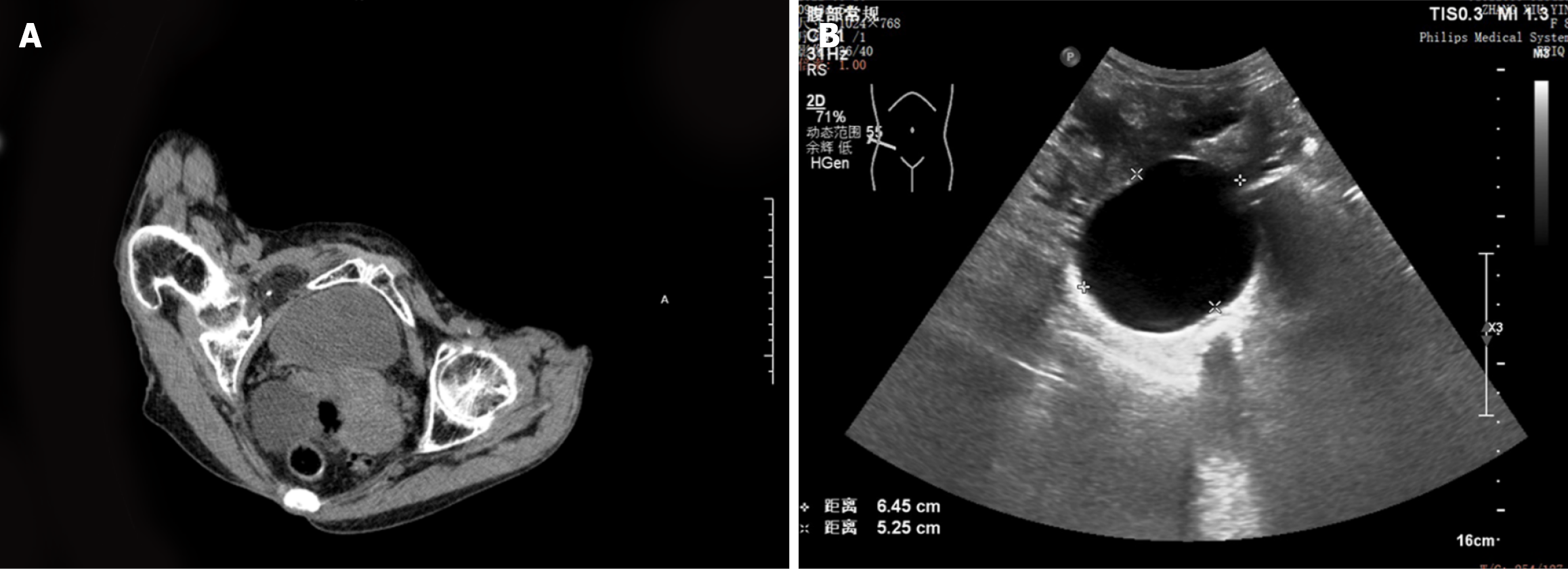
Figure 1 Preoperative imaging findings.
A: Computed tomography shows a round-like hypodense shadow outside the right obturator foramen, with punctate dense shadows inside; B: Abdominal color Doppler ultrasound shows a 65 mm × 52 mm cystic mass detected beside the right obturator externus muscle.
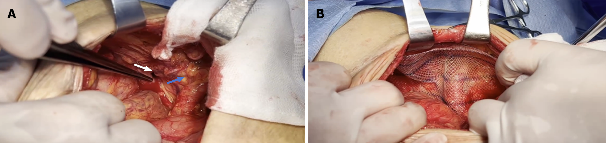
Figure 2 Intraoperative images.
A: Obturator foramen (white arrow), femoral canal (blue arrow); B: A hernia repair mesh was placed in the preperitoneal space.
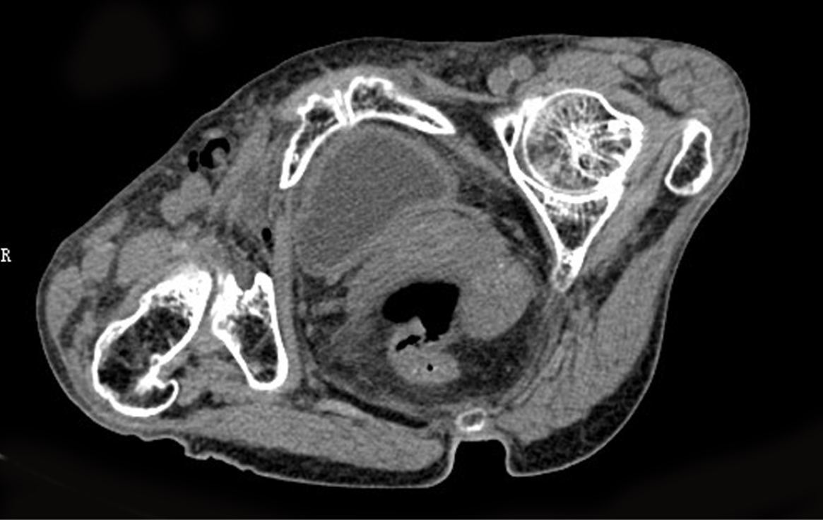
Figure 3 Abdominal computed tomography images of postoperative re-examination.
Imaging manifestations after repair of right obturator hernia combined with femoral hernia.
- Citation: Wang YF, Chen J, Wu LY. Obturator hernia combined with femoral hernia: A case report. World J Gastrointest Surg 2026; 18(5): 118072
- URL: https://www.wjgnet.com/1948-9366/full/v18/i5/118072.htm
- DOI: https://dx.doi.org/10.4240/wjgs.v18.i5.118072