
















Copyright: ©Author(s) 2026.
World J Gastrointest Surg. May 27, 2026; 18(5): 117647
Published online May 27, 2026. doi: 10.4240/wjgs.v18.i5.117647
Published online May 27, 2026. doi: 10.4240/wjgs.v18.i5.117647
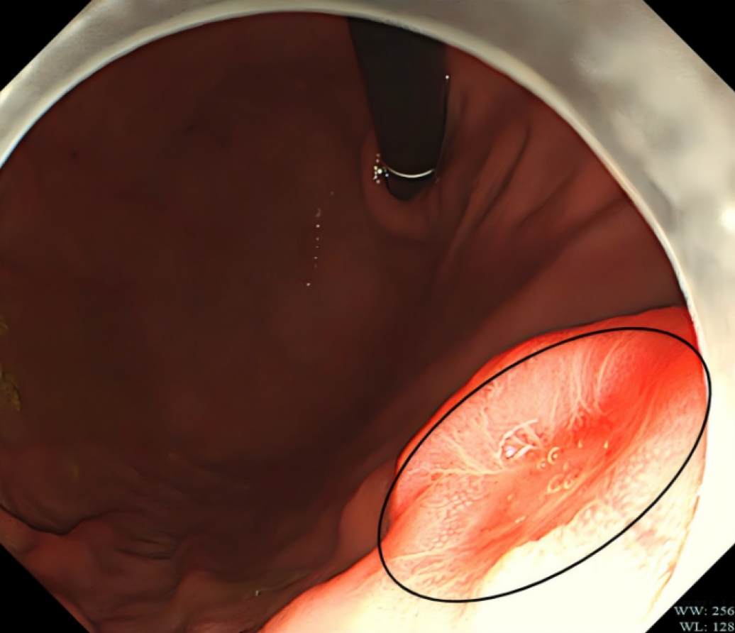
Figure 1
Lesion in the middle gastric body under gastroscopy (black circle).
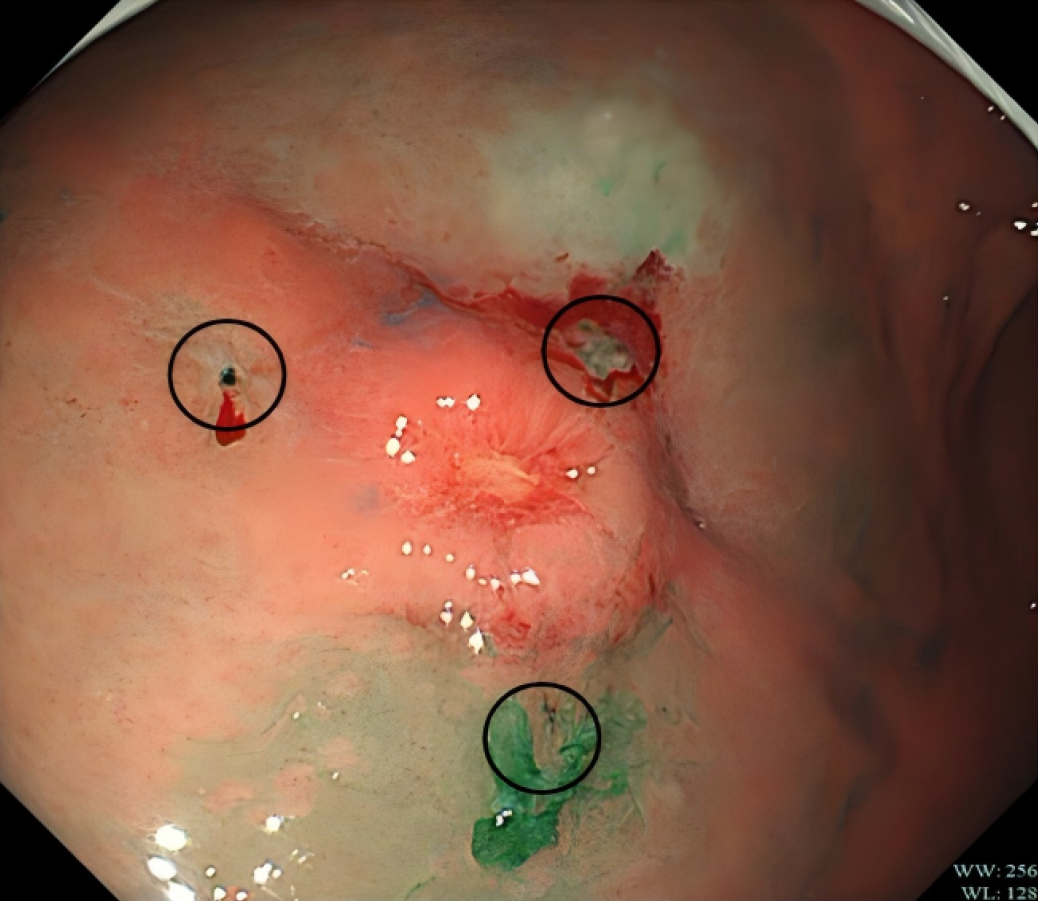
Figure 2
Injection of indocyanine green in the margin of the tumor (three black circles).
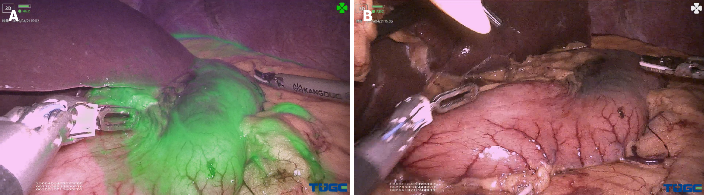
Figure 3 Tumor area in fluorescent mode and normal mode.
A: Fluorescent mode; B: Normal mode.
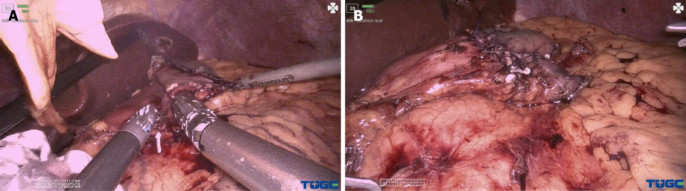
Figure 4 Digestive tract reconstruction.
A: Side-to-side anastomosis by linear cutter; B: Suture of common opening.
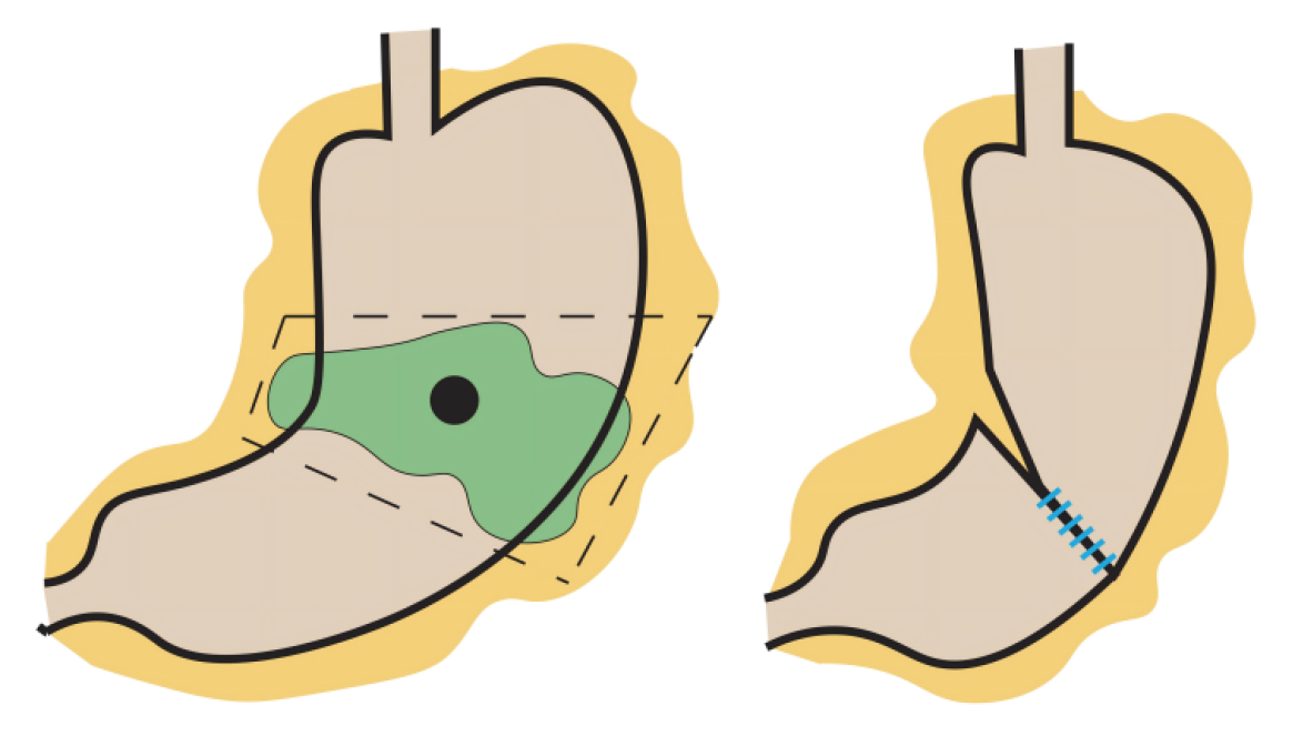
Figure 5
Brief schematic drawing of the intervention.
- Citation: Tou LZ, Wu D, Jiang C, Zheng JJ, Sun Z, Li JH, Que HF, Luo DS, Xu HT, Beisani M. Safety and efficacy of indocyanine green tracer-guided robotic segmental gastrectomy in early gastric cancer: A case report. World J Gastrointest Surg 2026; 18(5): 117647
- URL: https://www.wjgnet.com/1948-9366/full/v18/i5/117647.htm
- DOI: https://dx.doi.org/10.4240/wjgs.v18.i5.117647