
















Copyright: ©Author(s) 2026.
World J Gastrointest Surg. May 27, 2026; 18(5): 117633
Published online May 27, 2026. doi: 10.4240/wjgs.v18.i5.117633
Published online May 27, 2026. doi: 10.4240/wjgs.v18.i5.117633
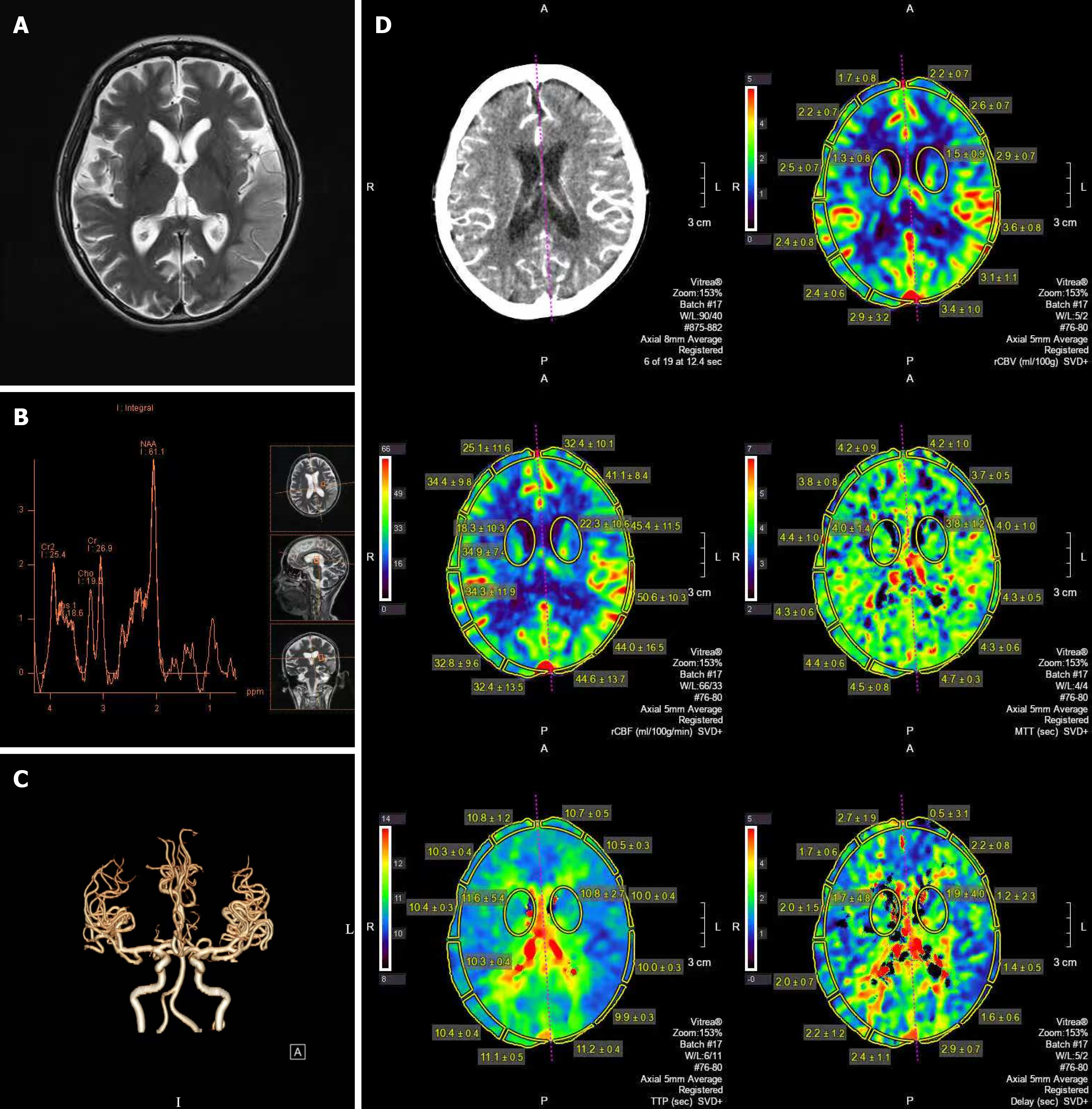
Figure 1 Magnetic resonance spectroscopy, cranial computed tomography angiography and cerebral computed tomography perfusion images.
A and B: Magnetic resonance spectroscopy showed patchy abnormal signal lesions in the left temporo-occipital lobe, and an elevated lactate peak was observed in the left temporal lobe; C and D: Cranial computed tomography angiography and cerebral computed tomography perfusion imaging showed no significant abnormalities.
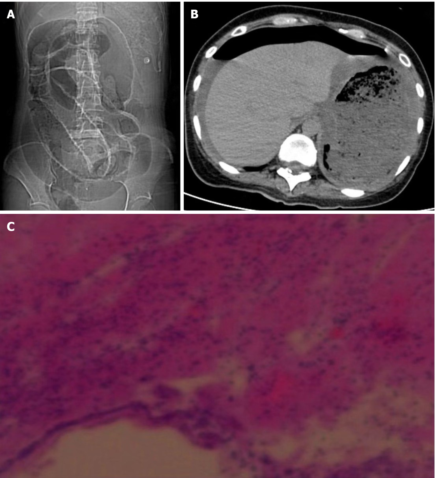
Figure 2 Whole abdominal computed tomography scan findings and postoperative histological examination.
A and B: Perforation of a hollow organ, diffuse thickening of the gastric wall; C: Loss of the superficial mucosal layer in the gastric wall tissue, accompanied by suppurative inflammation.
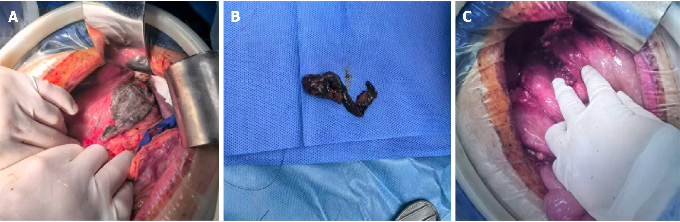
Figure 3 Intraoperative view.
A: A 10 cm laceration extending from the gastric body to the anterior wall of the gastric fundus presented; B: Resected black gangrene surrounding the gastric rupture; C: The gastric rupture was repaired.
- Citation: Zhang ZX, Jin BW, Wang S, Tao KL. Massive gastric perforation in a patient with mitochondrial encephalomyopathy, lactic acidosis and stroke-like episodes: A case report. World J Gastrointest Surg 2026; 18(5): 117633
- URL: https://www.wjgnet.com/1948-9366/full/v18/i5/117633.htm
- DOI: https://dx.doi.org/10.4240/wjgs.v18.i5.117633