
















Copyright: ©Author(s) 2026.
World J Gastrointest Surg. May 27, 2026; 18(5): 117565
Published online May 27, 2026. doi: 10.4240/wjgs.v18.i5.117565
Published online May 27, 2026. doi: 10.4240/wjgs.v18.i5.117565
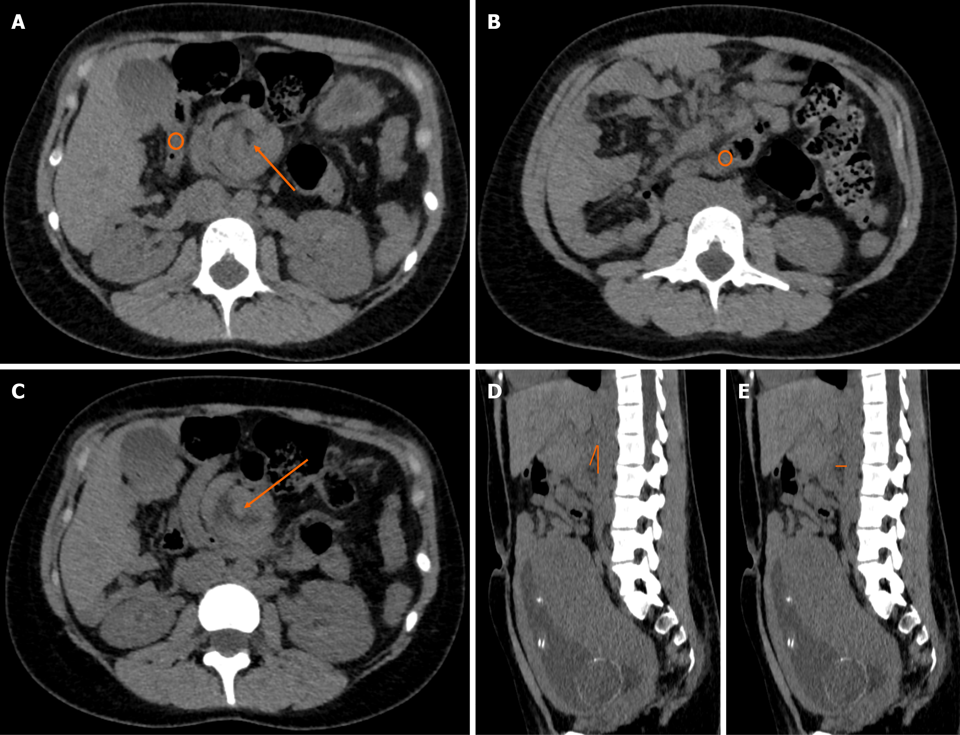
Figure 1 Non-contrast abdominal computed tomography in a 21-year-old primigravida demonstrating primary aortomesenteric (Wilkie’s) duodenal compression with a concomitant right paraduodenal hernia.
A-C: Axial images demonstrate a right paraduodenal hernia through Waldeyer’s fossa (orange arrow in A). The ascending colon and part of the hepatic flexure are contained within the hernia defect (orange circles in A and B). This anatomic distortion alters the course of the superior mesenteric artery, which is visualized centrally within the hernia defect (orange arrow in C). The third portion of the duodenum is poorly delineated and compressed at the aortomesenteric crossing, indicating that the duodenal obstruction arises from primary aortomesenteric compression rather than from the hernia itself; D and E: Sagittal reconstructions demonstrate a reduced aortomesenteric angle of approximately 19° (orange angle marker in D) and a reduced aortomesenteric distance of approximately 8 mm (orange arrow indicating the measurement in E). Because this pregnancy-adapted study lacked oral and intravenous contrast, these measurements are illustrative rather than diagnostic and are included to highlight the limitations of non-contrast computed tomography in pregnancy. Definitive confirmation of Wilkie’s syndrome was established intraoperatively.
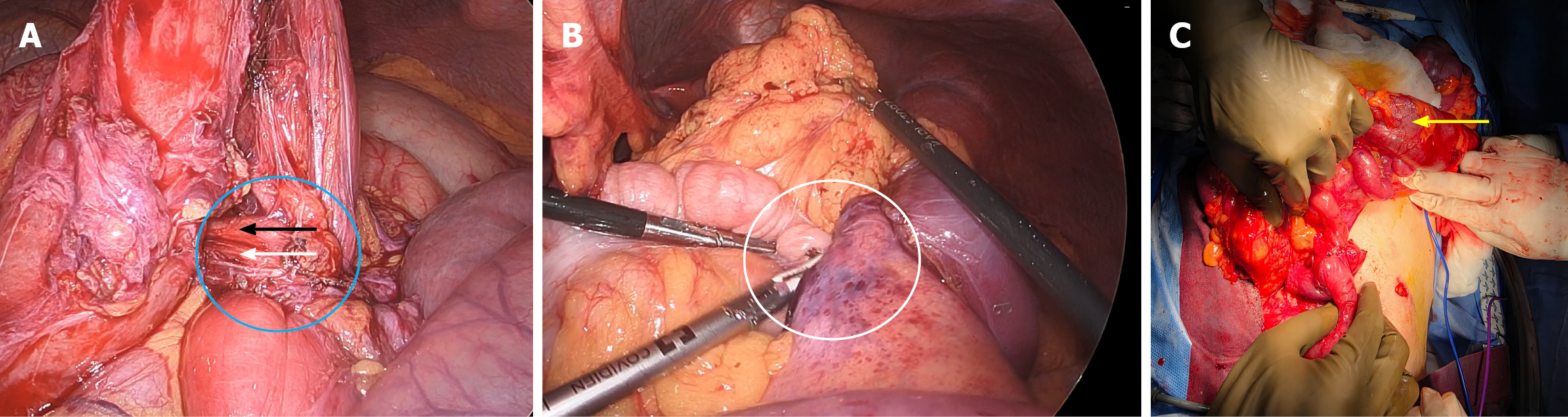
Figure 2 Intraoperative sequence demonstrating laparoscopic confirmation of aortomesenteric duodenal compression, identification of a concomitant right paraduodenal defect, and definitive reconstruction.
A: Laparoscopic view showing the third portion of the duodenum (blue circle) passing beneath the superior mesenteric artery (black arrow) and the superior mesenteric vein (white arrow), with extrinsic aortomesenteric compression causing focal luminal narrowing of third portion of the duodenum at the crossing; B: Laparoscopic view demonstrating the right paraduodenal (Waldeyer) defect, occupied by herniated transverse colon with adjacent bowel loops (white circle), such that the defect itself is not directly visualized because it is filled by the herniated intestinal contents; C: Open view following conversion to laparotomy, showing the completed side-to-side duodeno-duodenostomy (yellow arrow) restoring duodenal continuity.
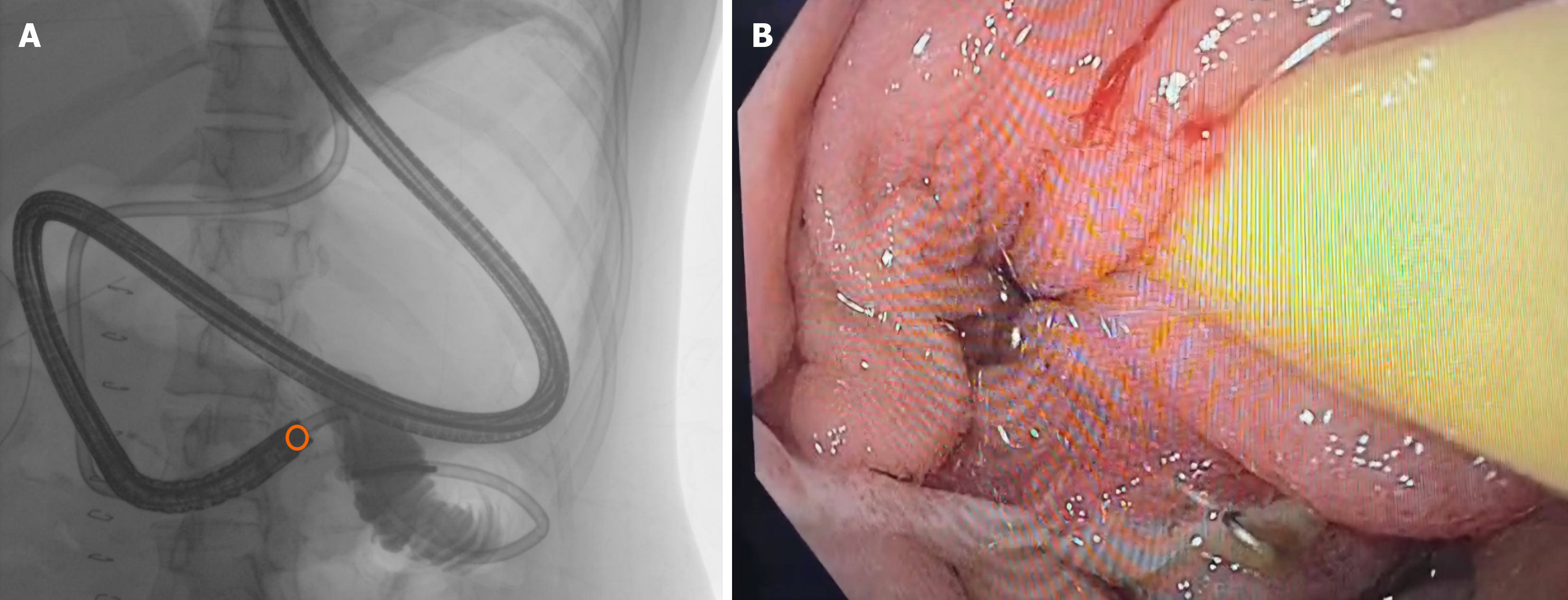
Figure 3 Postoperative assessment for transient gastroparesis demonstrating anastomotic patency.
A: Duodenoscopy in the operating room under real-time fluoroscopy shows advancement of the scope to the third portion of the duodenum (orange circle) with instillation of water-soluble contrast; no leak or collection is identified, and contrast passes freely into the proximal jejunum; B: Endoluminal duodenoscopic view demonstrating a widely patent side-to-side duodeno-duodenostomy with a nasojejunal tube traversing the anastomotic lumen.
- Citation: Sacasa F, Ploneda C, Cervantes M, Betancourt B, Casal J, Bedoya J, Valladares W. Superior mesenteric artery syndrome and right paraduodenal hernia causing intestinal obstruction during pregnancy: A case report. World J Gastrointest Surg 2026; 18(5): 117565
- URL: https://www.wjgnet.com/1948-9366/full/v18/i5/117565.htm
- DOI: https://dx.doi.org/10.4240/wjgs.v18.i5.117565