
















Copyright: ©Author(s) 2026.
World J Gastrointest Surg. May 27, 2026; 18(5): 116359
Published online May 27, 2026. doi: 10.4240/wjgs.v18.i5.116359
Published online May 27, 2026. doi: 10.4240/wjgs.v18.i5.116359
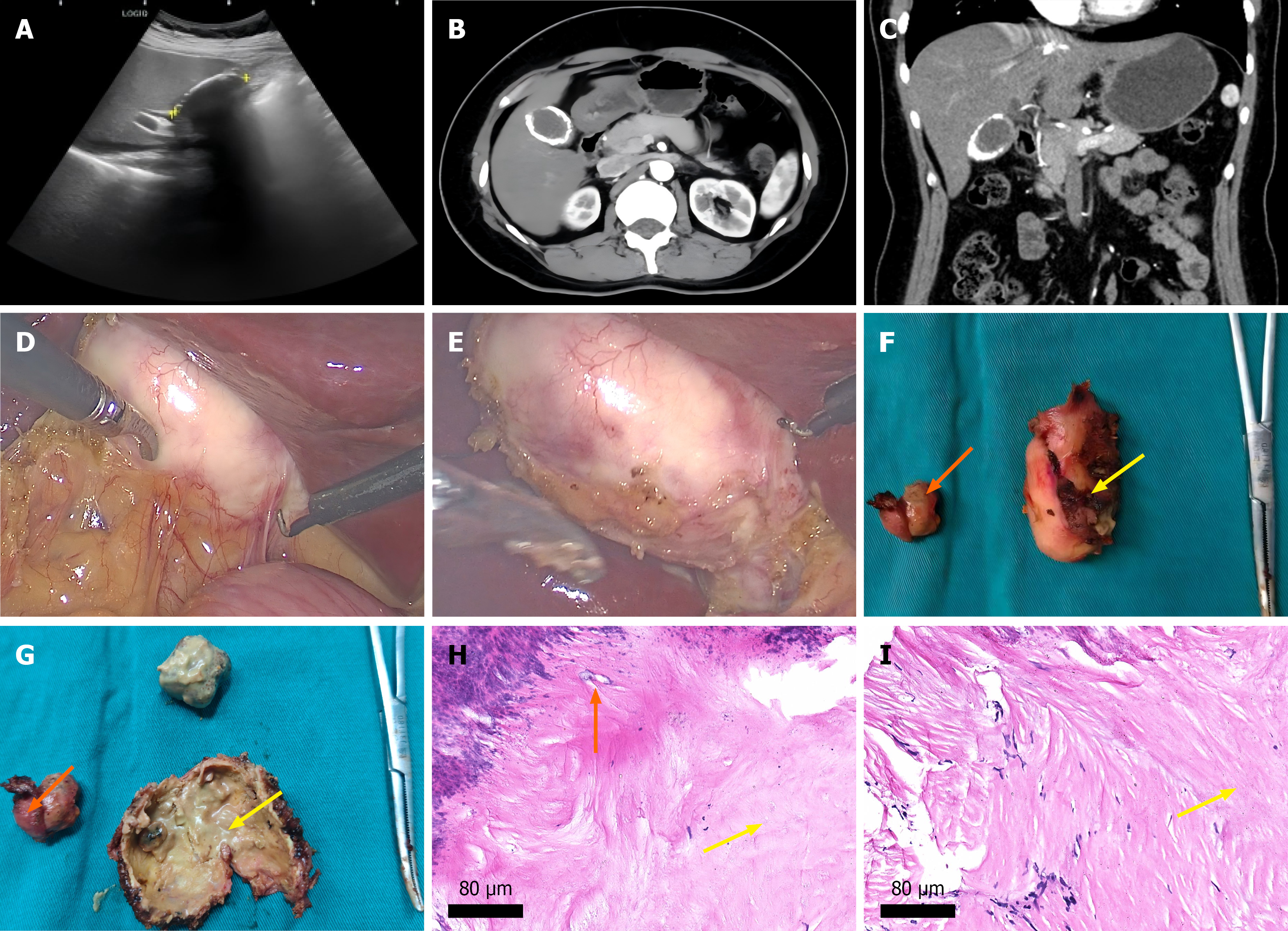
Figure 1 Imaging, intraoperative, and pathological findings of porcelain gallbladder.
A: Abdominal ultrasonography; B and C: Sagittal and coronal views of contrast-enhanced abdominal computed tomography; D and E: Laparoscopic intraoperative views; F and G: Resected gallbladder specimen (yellow arrows: Gallbladder body; orange arrows: Gallbladder infundibulum); H and I: Histological examination with hematoxylin-eosin staining (yellow arrows: Calcification foci; orange arrows: Hyaline degeneration).
- Citation: Chen SJ, Chen S, Hu AY, Li QS, Fan Y, Chen YG, Tian S, Li HL, Peng CJ, Han M. Clinical management of porcelain gallbladder: A case report. World J Gastrointest Surg 2026; 18(5): 116359
- URL: https://www.wjgnet.com/1948-9366/full/v18/i5/116359.htm
- DOI: https://dx.doi.org/10.4240/wjgs.v18.i5.116359