
















Copyright: ©Author(s) 2026.
World J Gastrointest Surg. May 27, 2026; 18(5): 115825
Published online May 27, 2026. doi: 10.4240/wjgs.v18.i5.115825
Published online May 27, 2026. doi: 10.4240/wjgs.v18.i5.115825
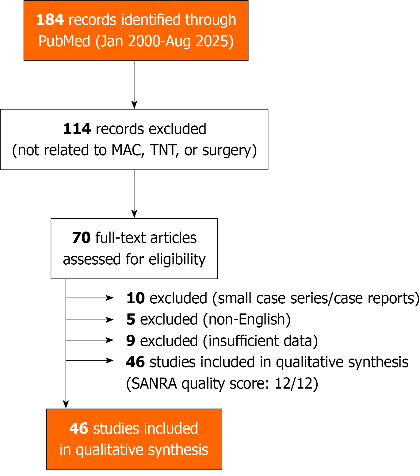
Figure 1 Article selection flow diagram.
The selection of studies included in the narrative review. From 184 records initially identified, 46 met the inclusion criteria, 44 clinical and surgical studies forming the main synthesis plus 2 additional articles separately addressing cost and resource utilization in mucinous adenocarcinoma (MAC) management. SANRA: Scale for the Assessment of Narrative Review Articles; TNT: Total neoadjuvant therapy.
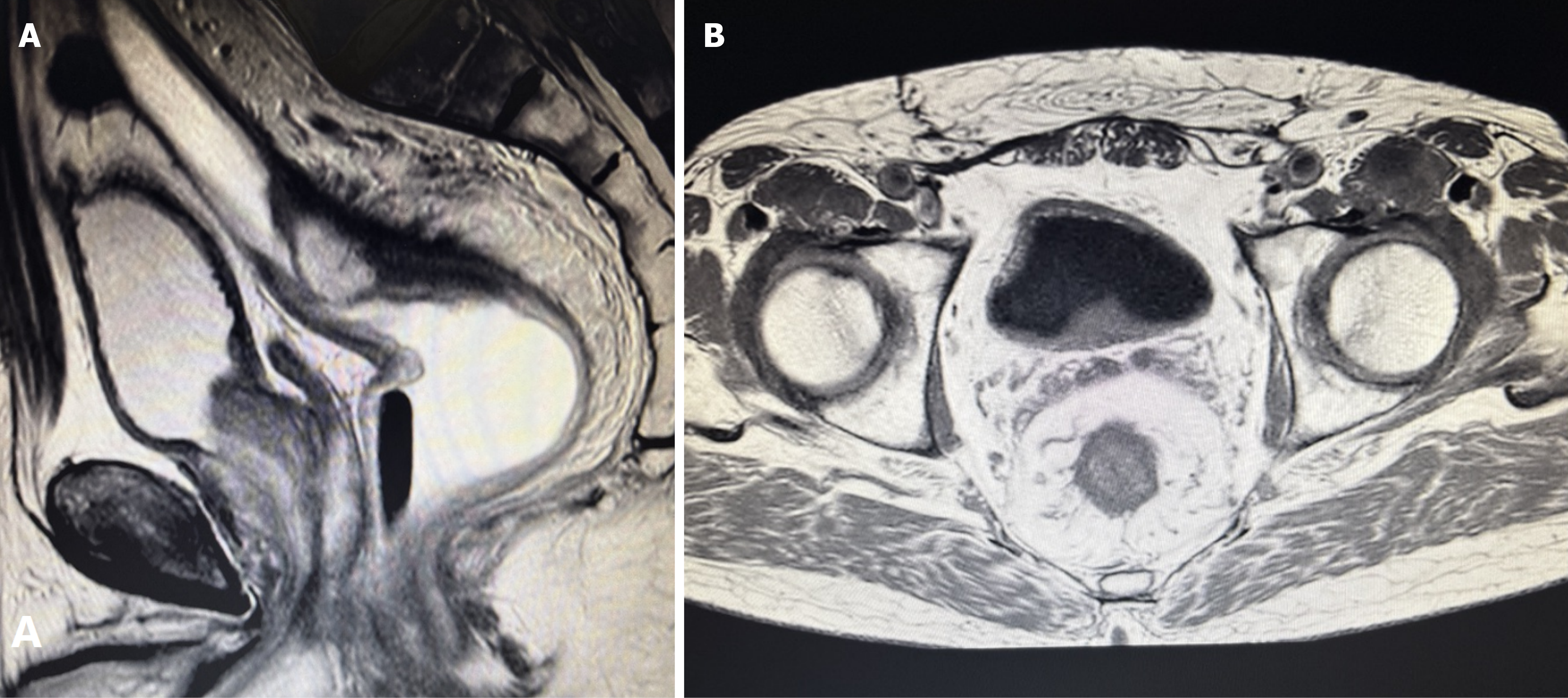
Figure 2 Pretreatment magnetic resonance imaging of a patient with mucinous adenocarcinoma.
A: Sagittal T2-weighted image showed a bulky, multilobulated rectal mass with markedly high signal intensity due to abundant extracellular mucin, extending toward the mesorectal fascia; B: Axial T2-weighted image demonstrated the same lesion with homogeneous hyperintense signal and poorly defined borders against the mesorectal fat, typical of mucinous tumors. These imaging features, high T2 signal, gelatinous internal texture, and ill-defined margins, are characteristic of mucinous histology and help differentiate mucinous adenocarcinoma from non-mucinous rectal adenocarcinoma.
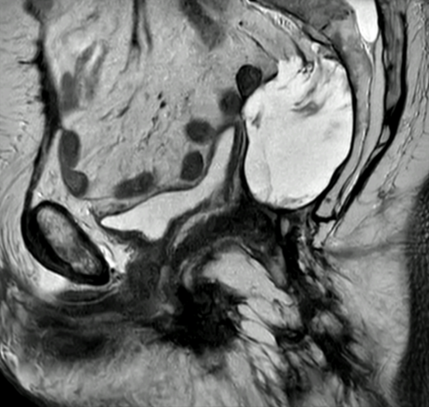
Figure 3 Pelvic magnetic resonance imaging revealed a local recurrence of mucinous adenocarcinoma 4 years subsequent to an R0 abdominoperineal resection.
Sagittal T2-weighted image demonstrated a lobulated, high-signal-intensity mass in the presacral space, consistent with recurrent mucinous disease. The lesion exhibited homogeneous T2 hyperintensity typical of extracellular mucin and ill-defined margins against adjacent pelvic structures. These imaging features are characteristic of mucinous local recurrence and help differentiate it from postoperative seroma or fibrosis.
- Citation: Patriti A, Graziano F, Chiari R. Mucinous rectal adenocarcinoma and neoadjuvant therapy: Implications for surgical practice. World J Gastrointest Surg 2026; 18(5): 115825
- URL: https://www.wjgnet.com/1948-9366/full/v18/i5/115825.htm
- DOI: https://dx.doi.org/10.4240/wjgs.v18.i5.115825