
















Copyright: ©Author(s) 2026.
World J Gastrointest Surg. Oct 27, 2026; 18(10): 123880
Published online Oct 27, 2026. doi: 10.4240/wjgs.123880
Published online Oct 27, 2026. doi: 10.4240/wjgs.123880
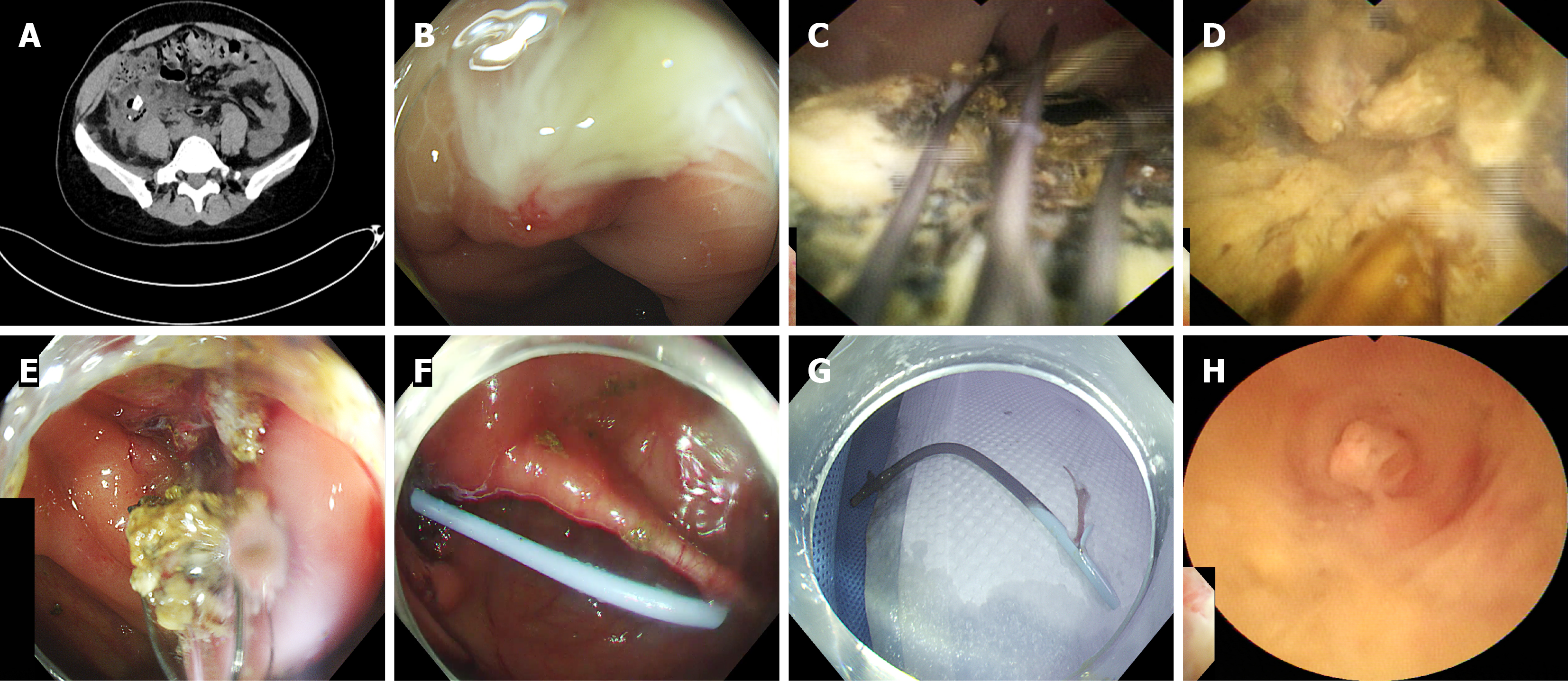
Figure 1 Representative computed tomography and endoscopic images of the patient across the treatment timeline.
A: Abdominal computed tomography revealed an abscess at the appendectomy site, containing a small amount of gas and a giant fecalith; B: Colonoscopy revealed a substantial amount of whitish purulent discharge adherent to the appendiceal orifice; C: Cholangioscopy revealed a large fecalith embedded within an abscess cavity adjacent to the appendiceal stump, and removal of the fecalith using a retrieval basket was unsuccessful; D: The electrohydraulic lithotripter successfully performed fecalith fragmentation; E: Most fragmented fecaliths were removed using a basket; F: A plastic stent was implanted into the abscess cavity; G: Two months later, the stent was removed via colonoscopy; H: Choledochoscopy enabled removal of the remaining few fecaliths, with marked improvement in the absorption of the abscess cavity.
- Citation: Li QL, Jiang B, Liu TY, Qin T. Endoscopic retrograde appendicitis therapy combined with electrohydraulic lithotripsy for stump appendicitis with abscess and fecalith: A case report. World J Gastrointest Surg 2026; 18(10): 123880
- URL: https://www.wjgnet.com/1948-9366/full/v18/i10/123880.htm
- DOI: https://dx.doi.org/10.4240/wjgs.123880