
















Copyright: ©Author(s) 2026.
World J Gastrointest Surg. Oct 27, 2026; 18(10): 123604
Published online Oct 27, 2026. doi: 10.4240/wjgs.123604
Published online Oct 27, 2026. doi: 10.4240/wjgs.123604
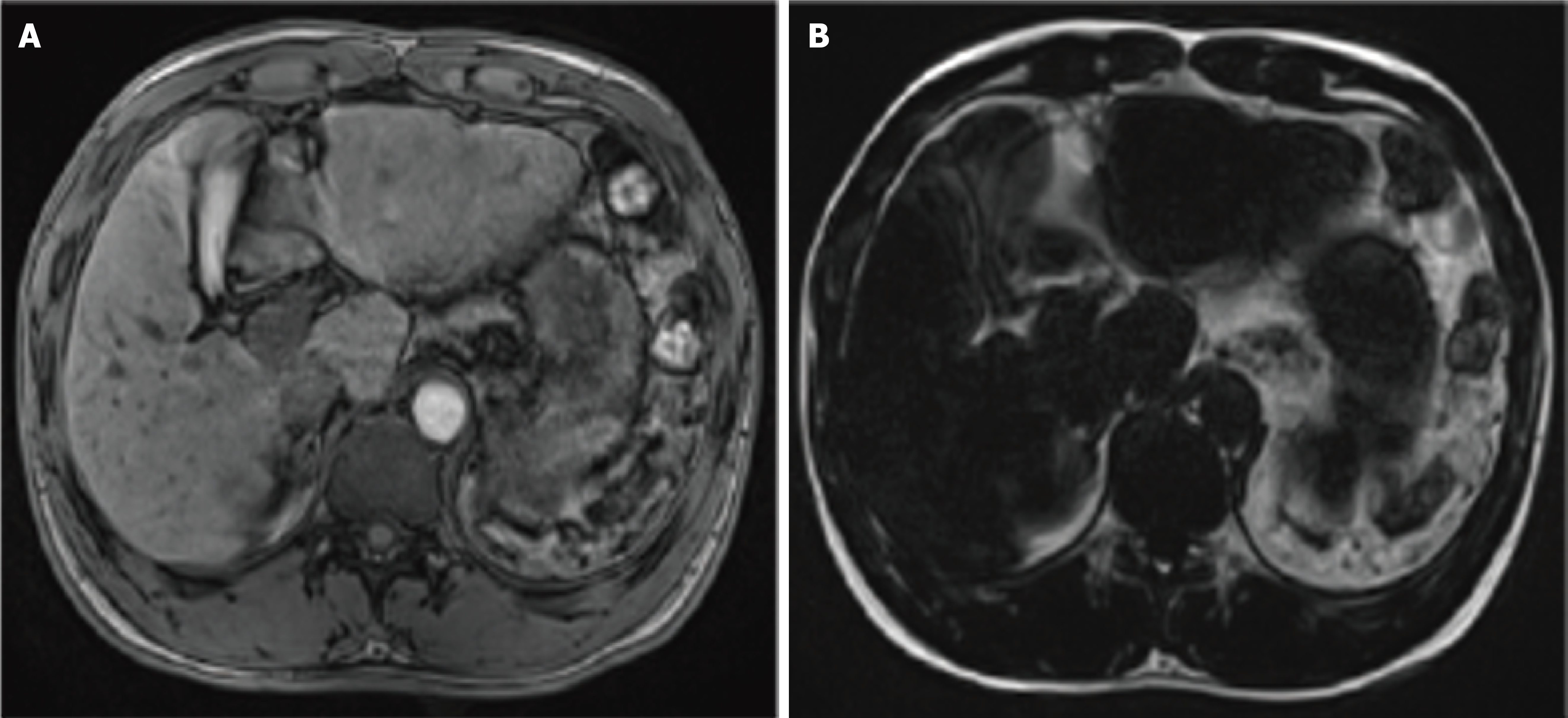
Figure 1 Magnetic resonance cholangiopancreatography findings in isolated congenital asplenia.
A: Axial T1-weighted image shows absence of the spleen, with no history of splenectomy; B: Axial T2-weighted image further confirms splenic agenesis, without evidence of accessory splenic tissue or other focal splenic lesions.
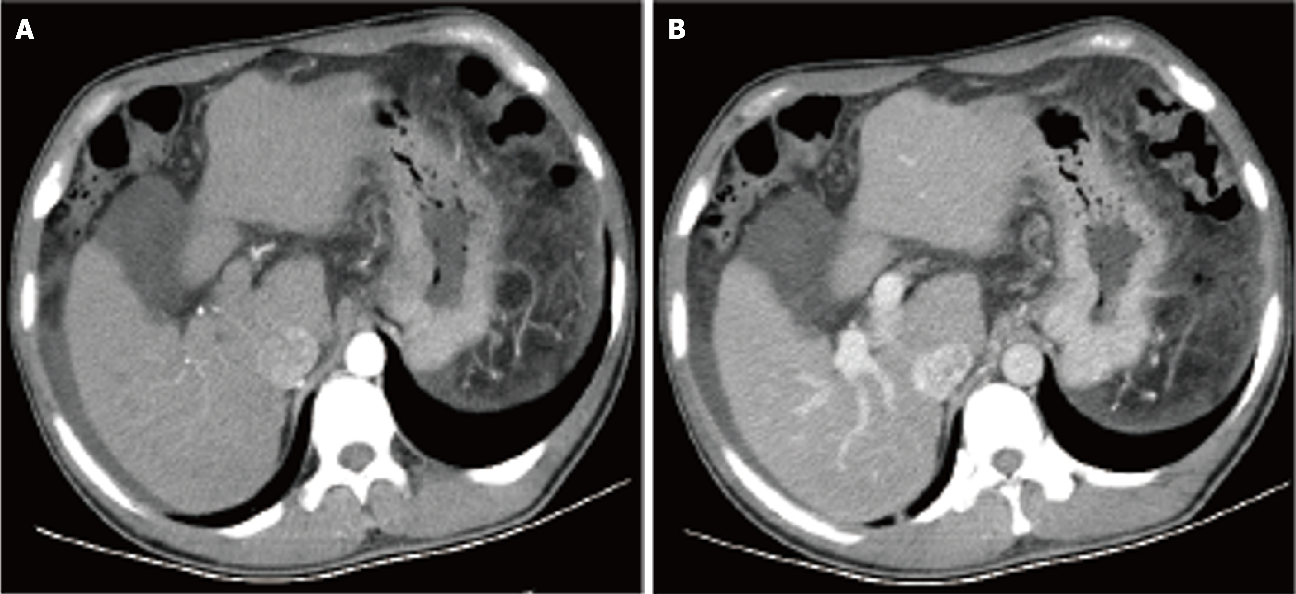
Figure 2 Portal vein computed tomography venography in isolated congenital asplenia.
A: Arterial phase computed tomography (CT) image demonstrates hepatic cirrhosis, absence of the spleen, and gallbladder wall edema with pericholecystic fluid; B: Venous phase CT image confirms portal hypertension, esophageal and gastric varices, and ascites, with no evidence of splenic tissue.
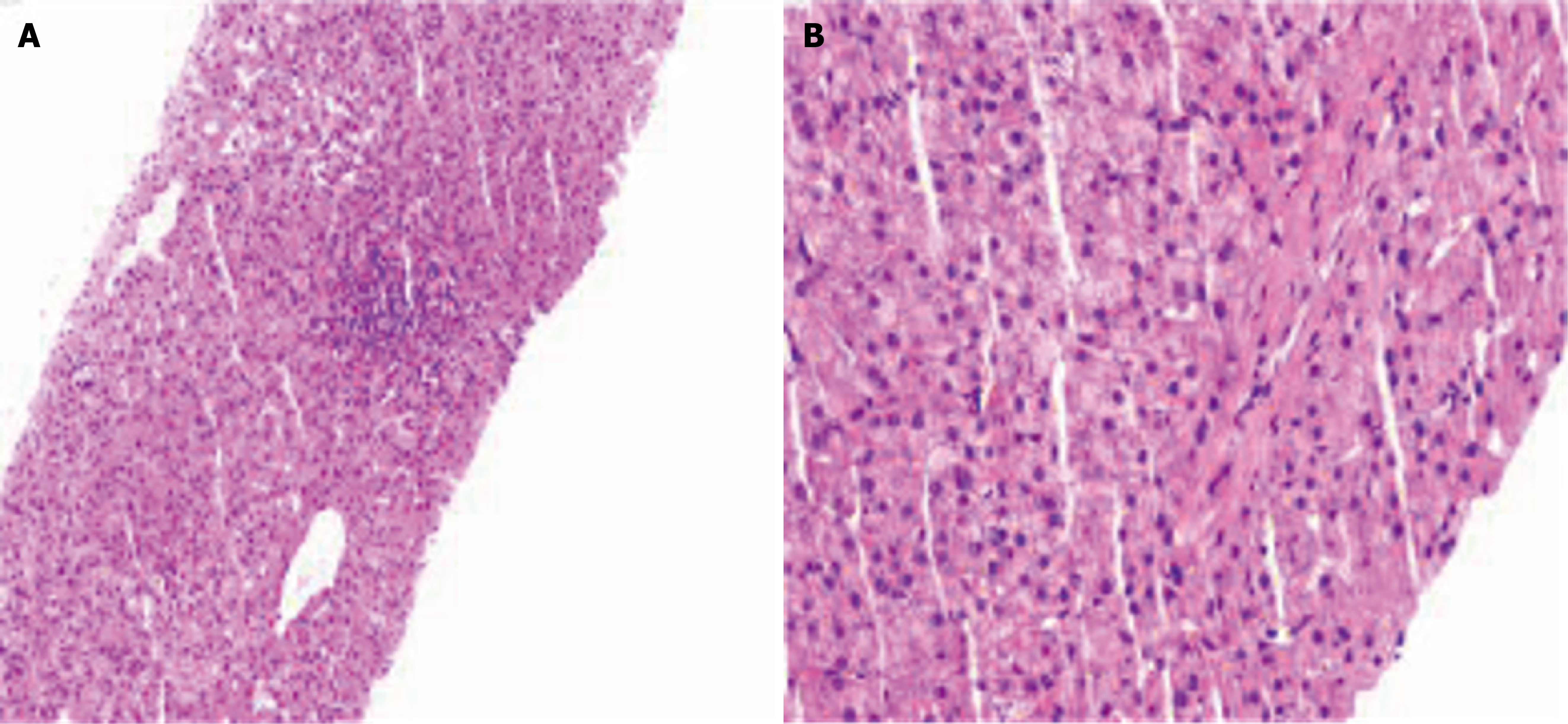
Figure 3 Pathological findings of ultrasound-guided liver biopsy.
A: Hematoxylin and eosin (H&E) staining (× 40); B: H&E staining (× 200).
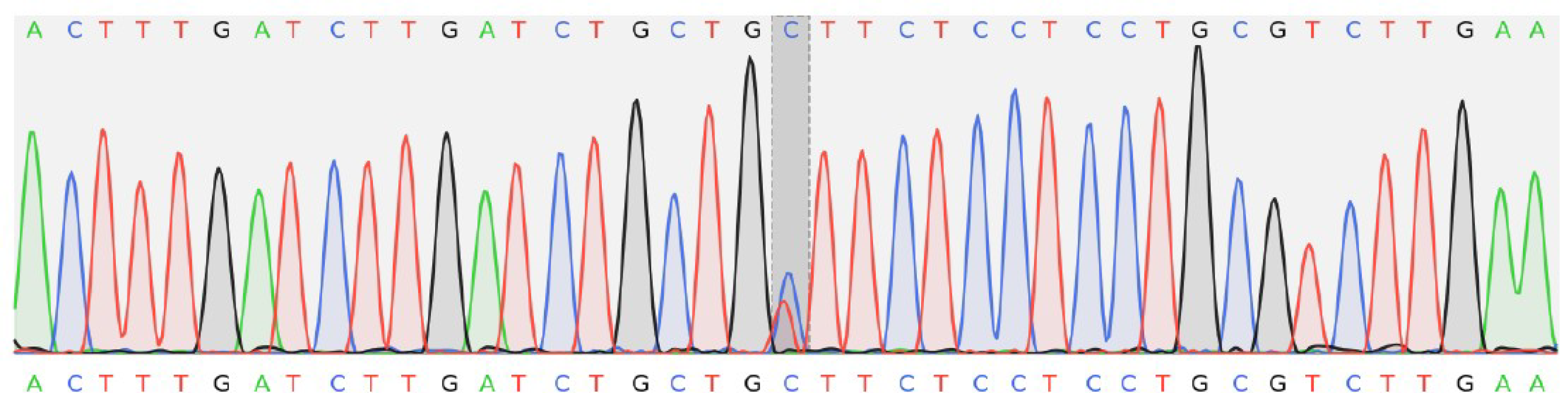
Figure 4 Genetic sequencing of a variant in the SON gene (chromosome 21 34927155, NM_138927.
1: C.5618G>A, p.Ser1873Asn).
- Citation: Zhang YY, Zhao YC, Chen LP, Yu HL. Isolated congenital asplenia with cryptogenic liver fibrosis and a novel SON variant: A case report. World J Gastrointest Surg 2026; 18(10): 123604
- URL: https://www.wjgnet.com/1948-9366/full/v18/i10/123604.htm
- DOI: https://dx.doi.org/10.4240/wjgs.123604