
















Copyright: ©Author(s) 2026.
World J Gastrointest Surg. Oct 27, 2026; 18(10): 123208
Published online Oct 27, 2026. doi: 10.4240/wjgs.123208
Published online Oct 27, 2026. doi: 10.4240/wjgs.123208
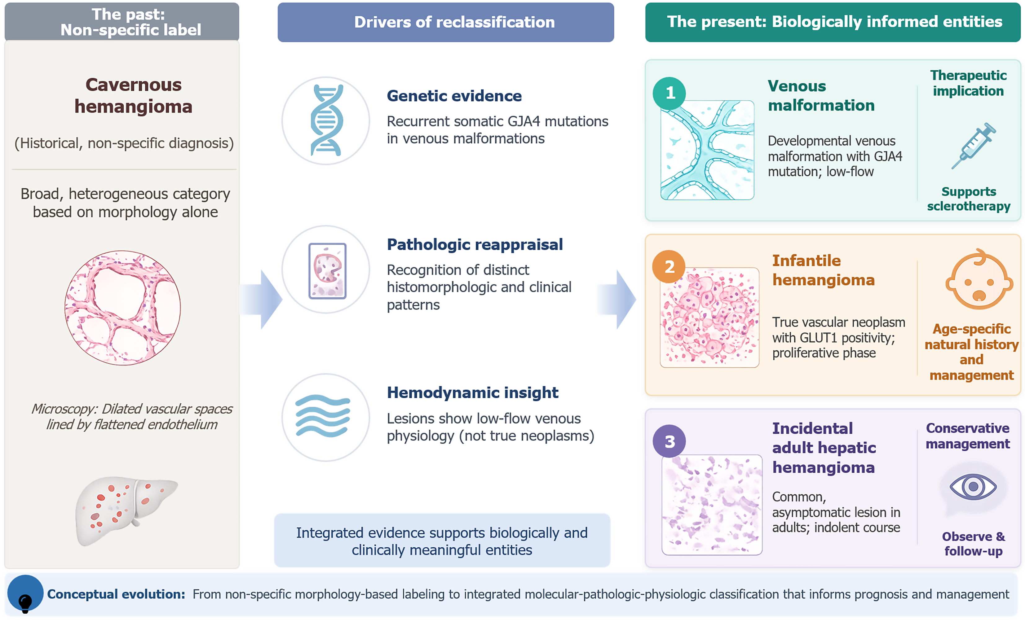
Figure 1 Emerging conceptual framework for adult hepatic vascular anomalies.
The figure contrasts the traditional umbrella term “cavernous hemangioma” with a biologically informed framework that recognizes possible overlap with low-flow venous malformation in selected adult lesions. This distinction may inform terminology, research classification, and selected interventional strategies. Gemini was used to assist in the design and visual optimization of the graphical abstract. The tool helped to optimize color palettes to better communicate the concepts visually. It was not used to generate or alter any empirical data.
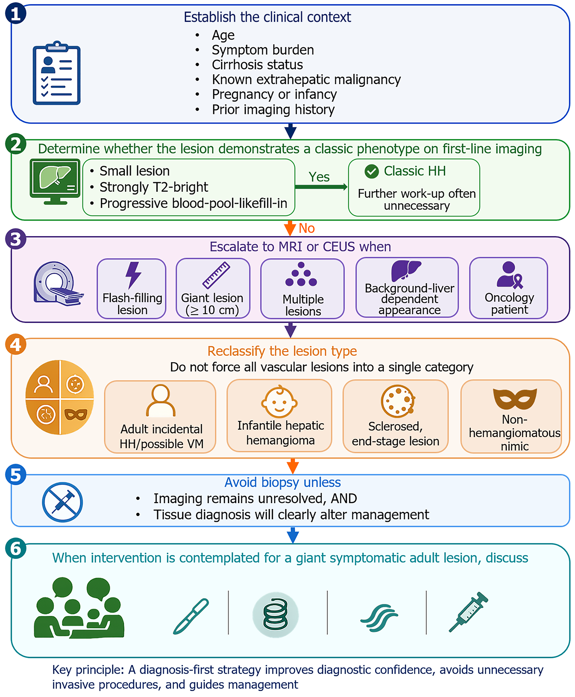
Figure 2 A practical diagnosis-first framework for hepatic hemangioma.
The flowchart illustrates a six-step multidisciplinary diagnostic and management strategy. The framework highlights the importance of clinical context (Step 1), specific indications for escalation to advanced multimodality imaging (Steps 2-3), the necessity of reclassifying lesion types (Step 4), stringent criteria for biopsy (Step 5), and customized interventional or surgical management based on lesion biology (Step 6). HH: Hepatic hemangioma; MRI: Magnetic resonance imaging; CEUS: Contrast-enhanced ultrasound; VM: Venous malformation.
- Citation: Ma YX, Zhou CM, Yilihaer Y, Jiang TM, Ruze R, Shao YM, Wen H, Aji T. Diagnosis and surgical decision-making in hepatic hemangioma: From venous malformation overlap to artificial intelligence-assisted multimodality imaging. World J Gastrointest Surg 2026; 18(10): 123208
- URL: https://www.wjgnet.com/1948-9366/full/v18/i10/123208.htm
- DOI: https://dx.doi.org/10.4240/wjgs.123208