
















Copyright: ©Author(s) 2026.
World J Gastrointest Surg. Oct 27, 2026; 18(10): 123036
Published online Oct 27, 2026. doi: 10.4240/wjgs.123036
Published online Oct 27, 2026. doi: 10.4240/wjgs.123036
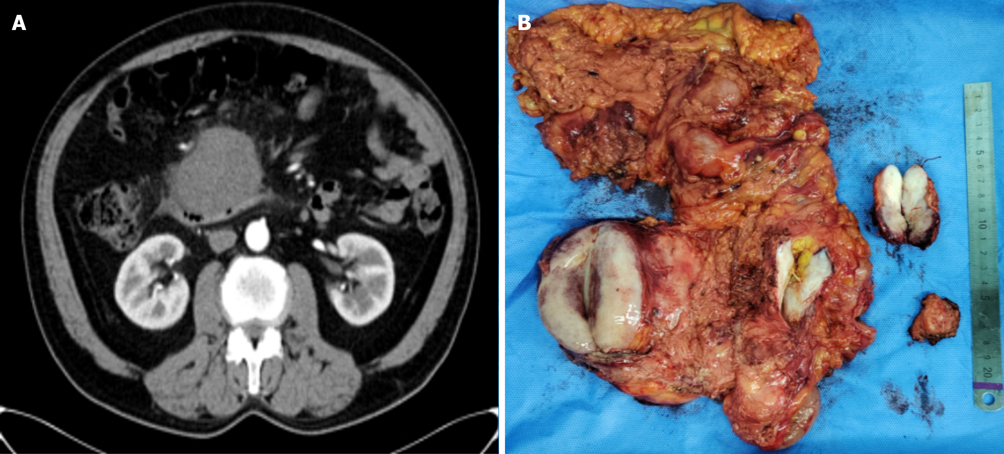
Figure 1 Preoperative imaging and resected specimen of the abdominal masses.
A: Preoperative enhanced computed tomography showing mesenteric tumor of the right hemicolon (image from the arterial phase); B: Resected specimen of the mesentery with multiple tumor nodules and the right hemicolon.
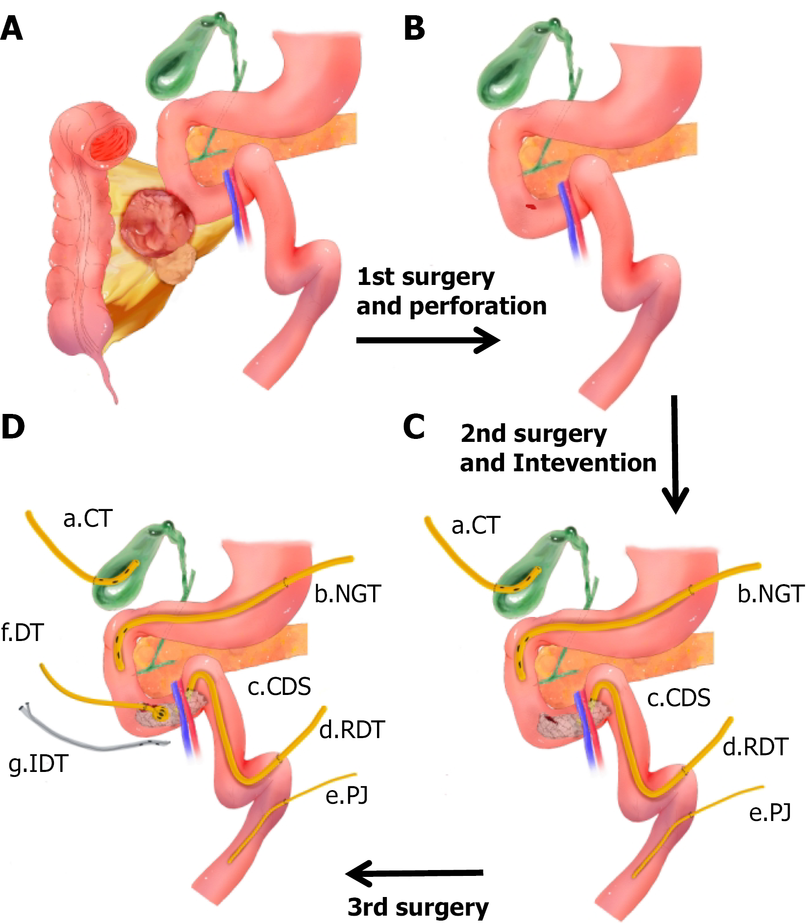
Figure 2 Surgical illustrations.
A and B: First operation: Multiple soft tissue tumors of the colonic mesentery and the right hemicolon were resected. The larger tumor was tightly adherent to the lateral wall of the duodenum; B and C: Second operation and intervention: Intraoperative exploration revealed a perforation on the anterior wall of the horizontal portion of the duodenum. The perforation was repaired with sutures and reinforced using a ligamentum teres hepatis patch. Gastroenteric and biliary diversions were created, and a feeding jejunostomy tube was placed. Postoperative examination indicated persistent unhealed duodenal perforation, and a covered duodenal stent was placed over the perforation site; C and D: Third operation: A duodenostomy tube was directly placed at the perforation site and secured with a wrap of the ligamentum teres hepatis. Additionally, a paraduodenal drain with irrigation and suction capabilities was placed. CT: Cholecystostomy tube; NT: Nasogastric tube; CDS: Ccovered duodenal stent; RDT: Retrograde duodenal decompression tube; PJ: Percutaneous jejunostomy tube; DT: Duodenostomy tube; IDT: Irrigable drainage tube.
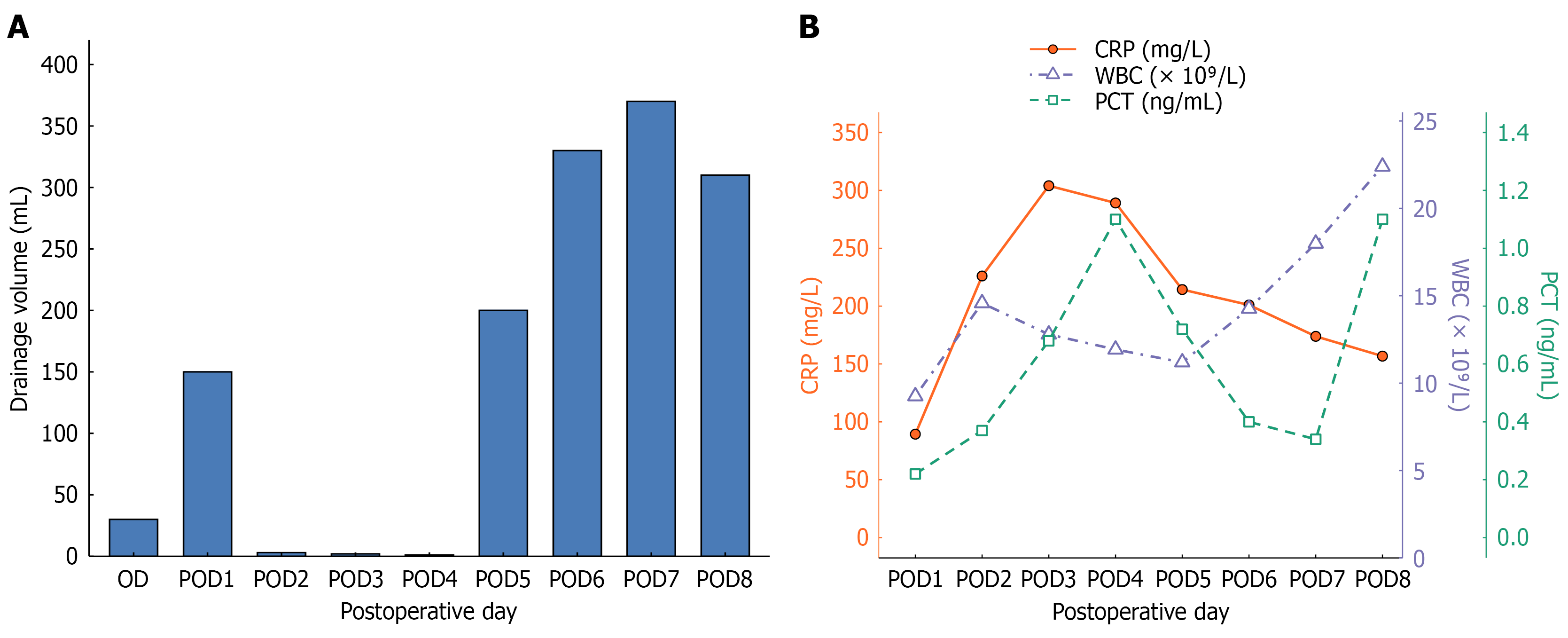
Figure 3 Postoperative drainage volume and inflammatory markers.
A: Daily drainage volume from the operation day through postoperative day 8; B: Serial serum C-reactive protein, white blood cell count, and procalcitonin measurements from postoperative day 1 to postoperative day 8. POD: Postoperative day; CRP: C-reactive protein; WBC: White blood cell; PCT: Procalcitonin.
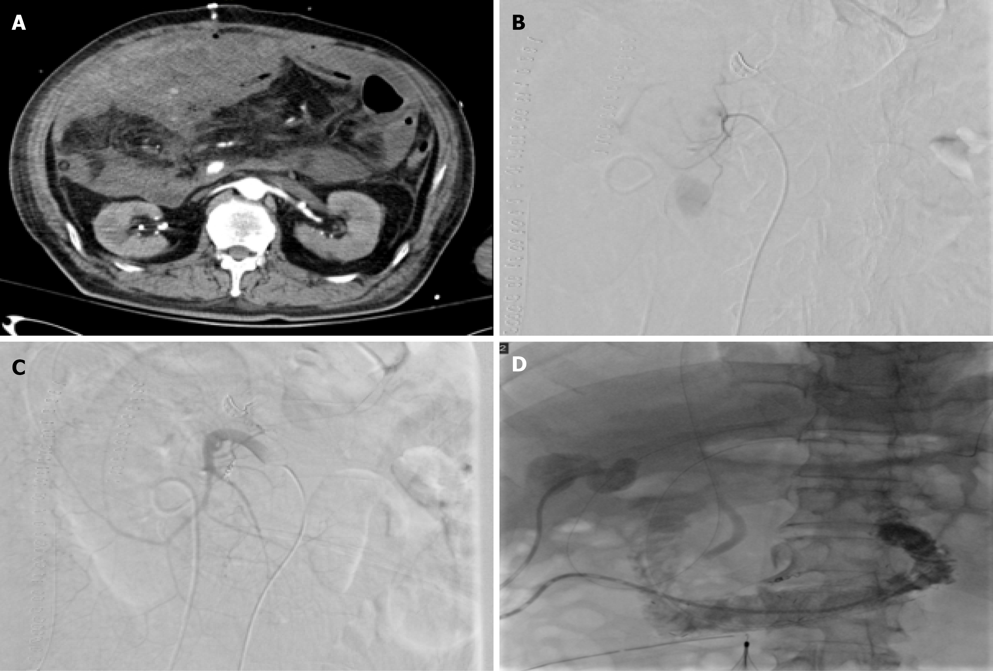
Figure 4 Imaging and interventional management of postoperative intra-abdominal hemorrhage and duodenal perforation.
A and B: Enhanced computed tomography and angiography showed contrast extravasation from abdominal vessels, suggesting active bleeding; C: Consequently, embolization of the superior mesenteric artery branches was performed; D: A covered stent was placed at the site of the duodenal perforation under interventional guidance.
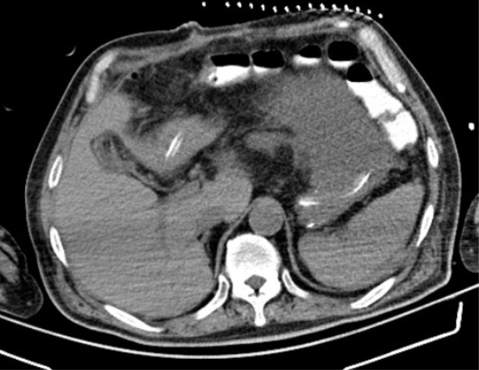
Figure 5 Targeted puncture drainage under interventional radiology guidance at two major sites of fluid collections adjacent to the stomach and within the abdominal cavity.
Beyond the direct manifestations of duodenal perforation, notably hemorrhage and infection, the patient’s recovery was complicated by a series of challenges secondary to extensive surgical interventions and the critical nature of his overall condition. The patient presented with extensive deep venous thrombosis, coagulopathy, urinary system dysfunction, malnutrition, and the need for rehabilitation and physical therapy. Under mul
- Citation: Dong LB, Wu MW, Han JS, Lin C, Wang WB. Multidisciplinary management of postoperative duodenal perforation using multiple drainage and stenting: A case report and review of literature. World J Gastrointest Surg 2026; 18(10): 123036
- URL: https://www.wjgnet.com/1948-9366/full/v18/i10/123036.htm
- DOI: https://dx.doi.org/10.4240/wjgs.123036