Copyright
©The Author(s) 2025.
World J Gastrointest Surg. Jul 27, 2025; 17(7): 106700
Published online Jul 27, 2025. doi: 10.4240/wjgs.v17.i7.106700
Published online Jul 27, 2025. doi: 10.4240/wjgs.v17.i7.106700
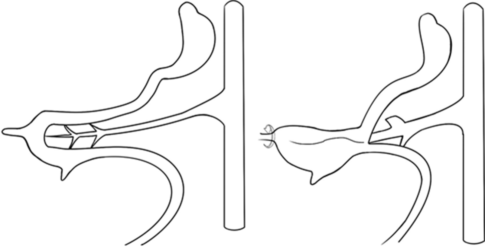
Figure 1 Intestinal rotation occurring in the 6th-8th weeks of embryonic development.
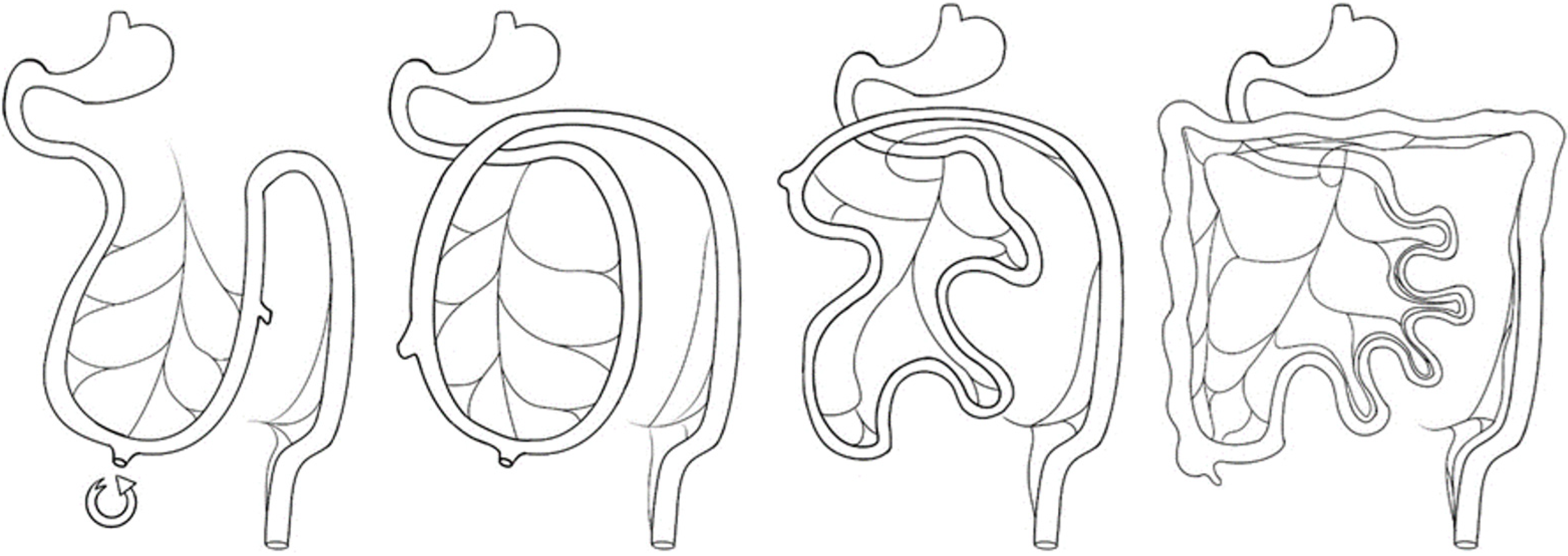
Figure 2 Normal rotation of the fetal intestinal tube.
Figure 3 Pre-operative and post-operative computed tomography imaging of duodenal and jejunal pathologies: Membranous wrapping and obstruction management.
A: Preoperative computed tomography showed that the duodenum and upper jejunum intestine were clustered and seemed to be wrapped with membrane (blue arrows). The orange arrow notes the transverse colon passing behind the small intestine; B and C: Computed tomography reexamination after surgery showed the horizontal segment of duodenum with an intestinal obstruction catheter (blue arrow). The orange arrow notes the transverse colon passing behind the duodenum.
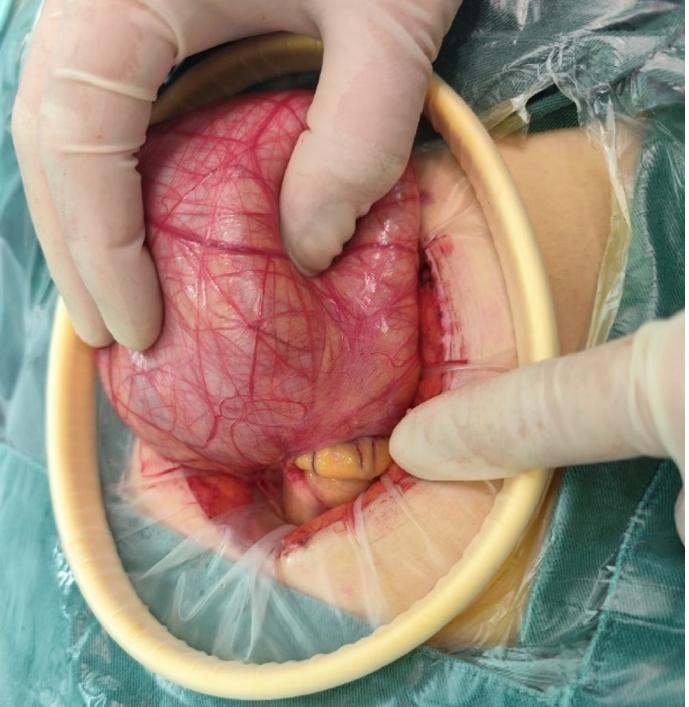
Figure 4 The superior jejunum is herniated by the omentum.
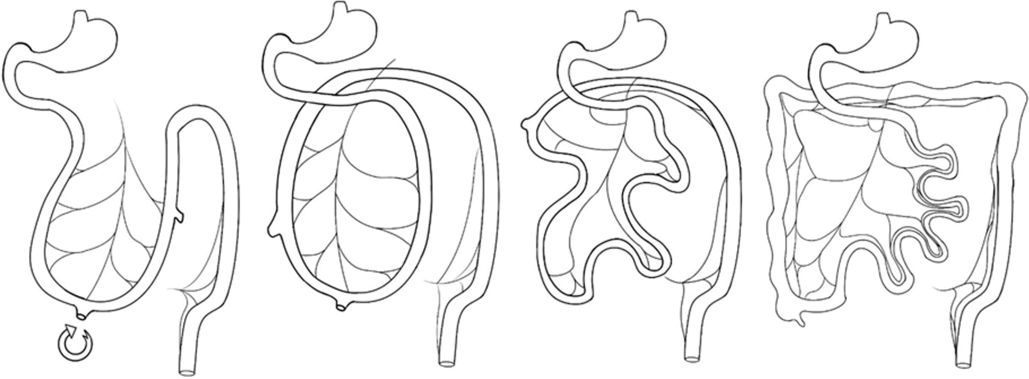
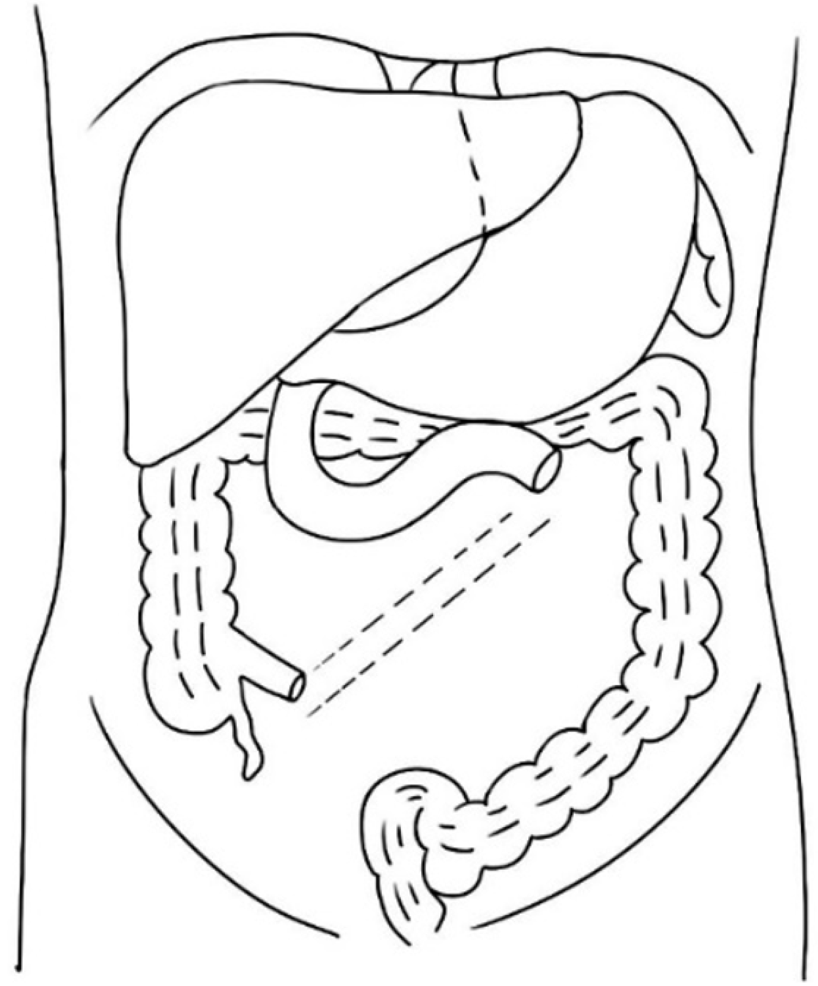
Figure 5 Congenital midgut reverse transposition.
Figure 6 Linear mesenteric root fixation.
- Citation: Wang Q, Sun K, Gong XS. Congenital midgut reverse transposition with herniation of the jejunum into a malformed omentum: A case report. World J Gastrointest Surg 2025; 17(7): 106700
- URL: https://www.wjgnet.com/1948-9366/full/v17/i7/106700.htm
- DOI: https://dx.doi.org/10.4240/wjgs.v17.i7.106700


