Copyright
©The Author(s) 2025.
World J Gastrointest Surg. Jul 27, 2025; 17(7): 106471
Published online Jul 27, 2025. doi: 10.4240/wjgs.v17.i7.106471
Published online Jul 27, 2025. doi: 10.4240/wjgs.v17.i7.106471
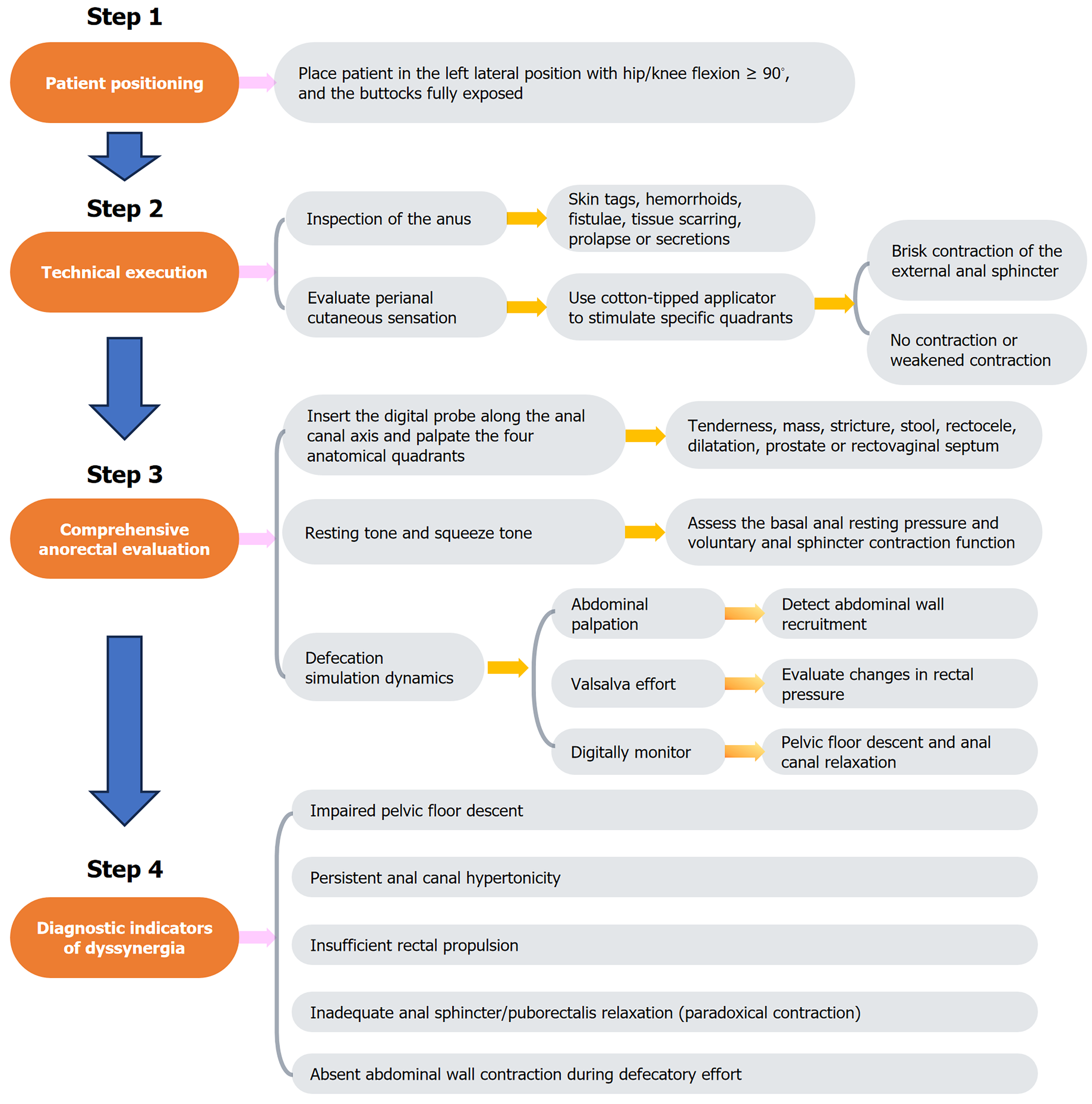
Figure 1 Steps of digital rectal examination.
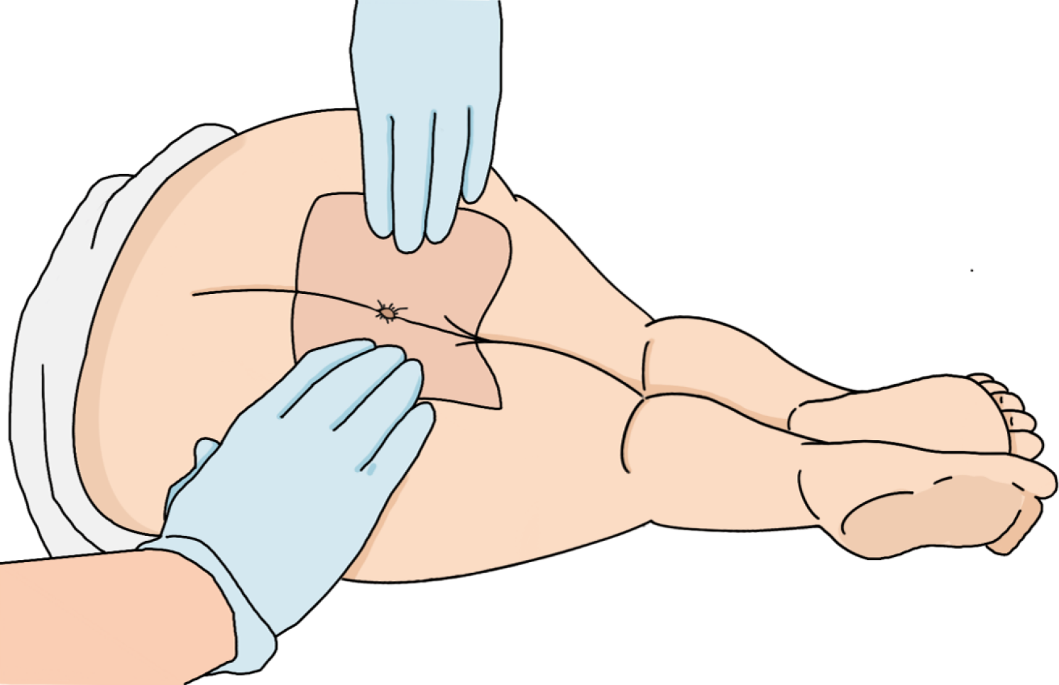
Figure 2 Patient positioning for digital rectal examination.
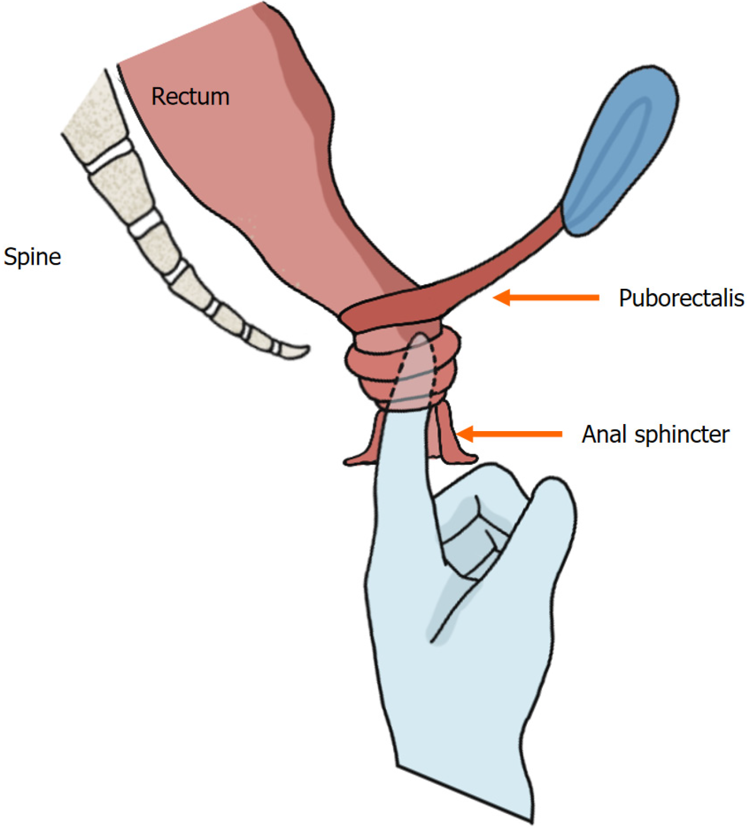
Figure 3 Digital insertion and anorectal palpation.
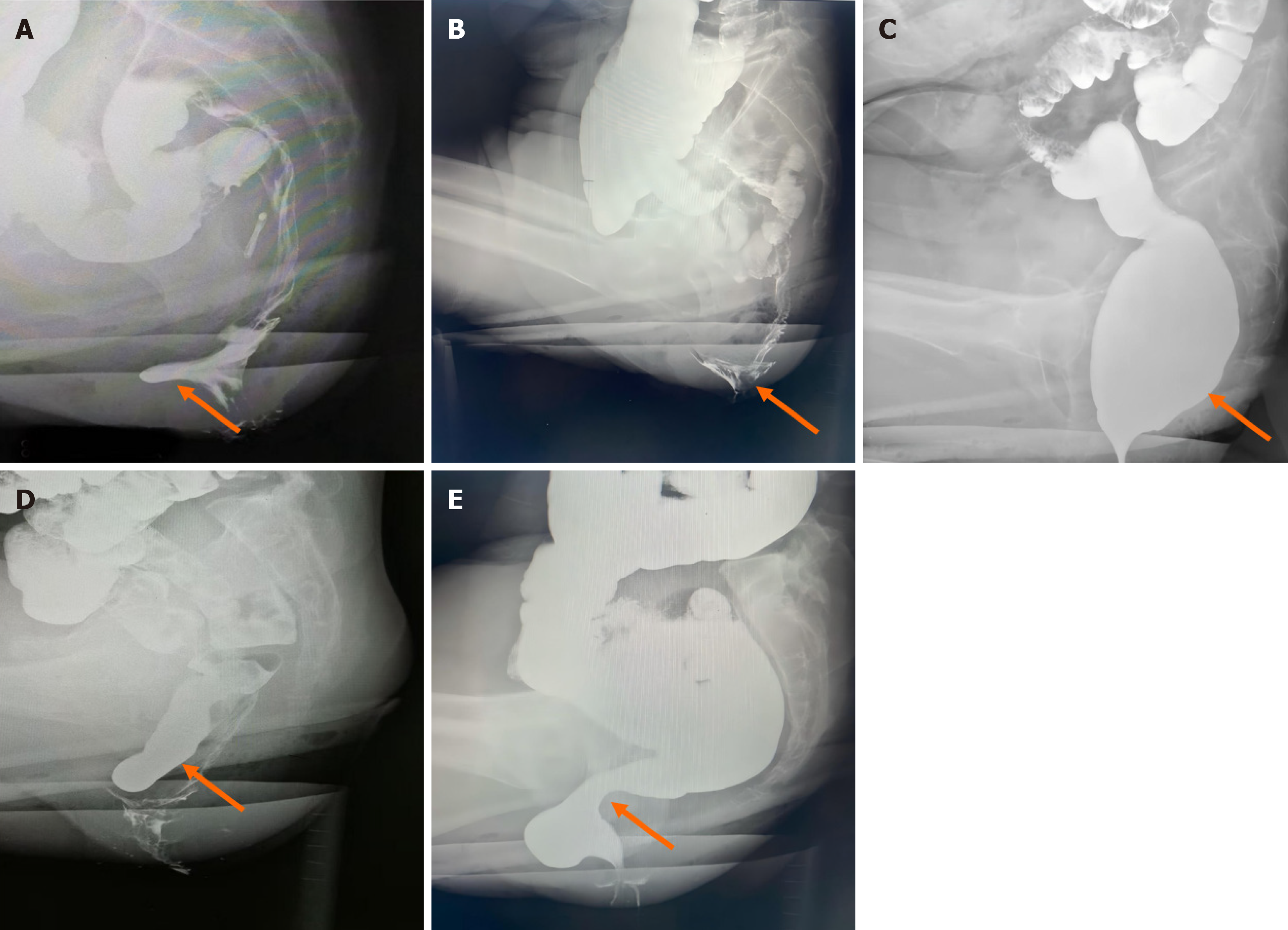
Figure 4 Morphological changes demonstrated by defecography (indicated by the arrow).
A: Sac-like protrusion of the anterior rectal wall; B: Internal rectal prolapse in a funnel-shaped configuration; C: Rectal luminal distension; D: The sigmoid colon descends below the pubococcygeal line; E: Evident pressure trace of the puborectalis muscle.
- Citation: Zhu LJ, Zeng XL, Yang XD. Enhancing clinical practice: The role of digital rectal examination in diagnosing functional defecation disorders. World J Gastrointest Surg 2025; 17(7): 106471
- URL: https://www.wjgnet.com/1948-9366/full/v17/i7/106471.htm
- DOI: https://dx.doi.org/10.4240/wjgs.v17.i7.106471


