Copyright
©The Author(s) 2025.
World J Gastrointest Surg. Jul 27, 2025; 17(7): 106365
Published online Jul 27, 2025. doi: 10.4240/wjgs.v17.i7.106365
Published online Jul 27, 2025. doi: 10.4240/wjgs.v17.i7.106365
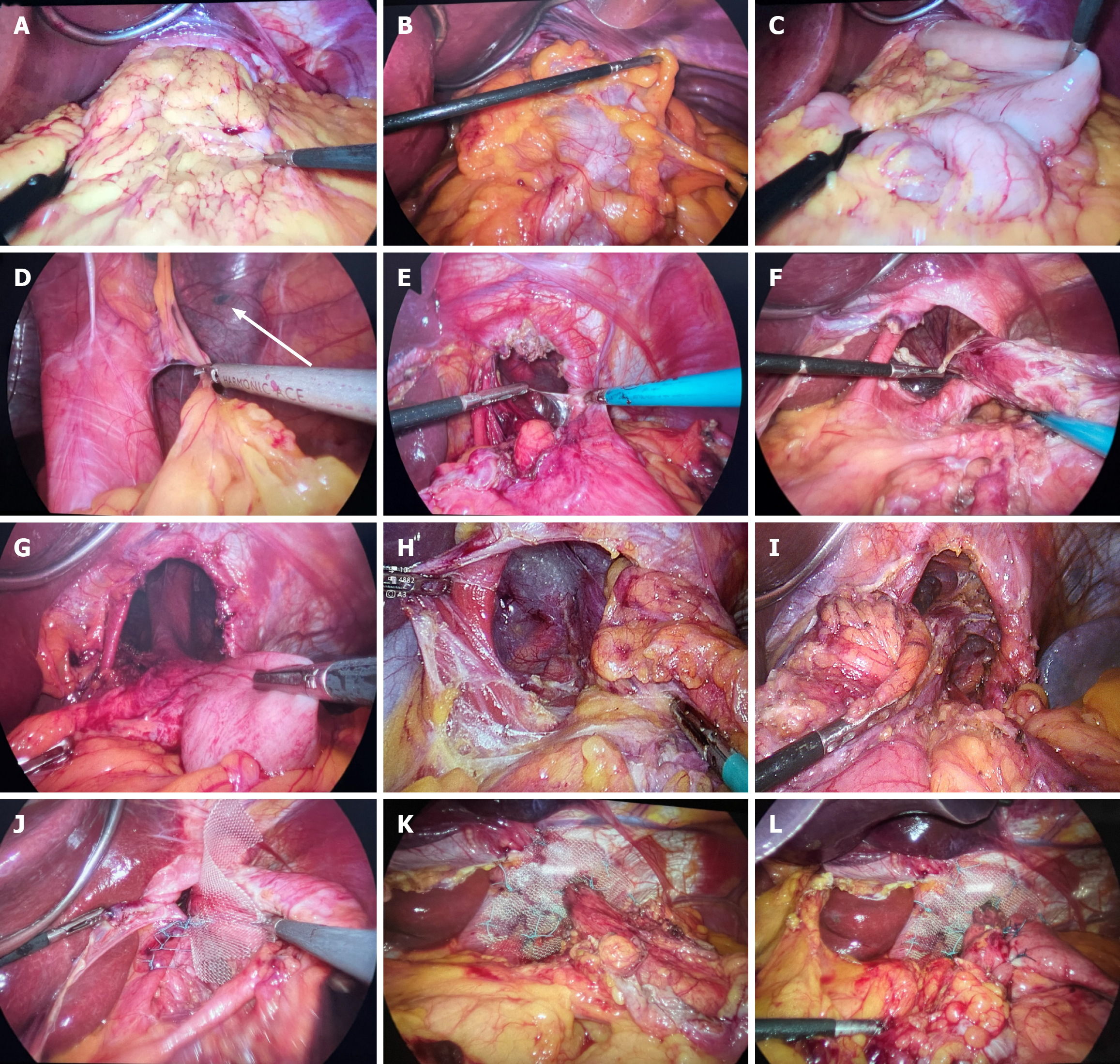
Figure 1 Surgical approach results.
A-D: Mobilization and complete evacuation of the hernia contents into the abdominal cavity (A: Type IV hiatal hernia; The esophageal hiatus is filled with great omentum and abdominal organs; B: Reduction of the transverse colon; C: Reduction of the stomach; D: Mobilization of the tissues inside the hernia sac in the mediastinum. The arrow shows the tissue of the left lung covered with the hernia sac); E-G: Mobilization of the neck of the hernia sac at the level of the diaphragmatic crura (E: A section of the neck of the hernia sac; F: Mobilization is performed, and the grasper fixes the edge of the hernia sac; G: The hernia sac is left in the mediastinum, and the neck of the sac is mobilized at the level of the diaphragmatic crura; H and I: Mobilization of the hernia sac); H: The hernia sac is mobilized and pulled down between the right crura and the esophagus; I: The hernia sac is mobilized and pulled down between the left crura and the esophagus; J-L: Reinforcement of the esophageal hiatus and fundoplication (J: Posterior crurorrhaphy was performed with nonabsorbable sutures; K: Polypropylene mesh was fixed around the esophagus; L: Nissen fundoplication was performed).
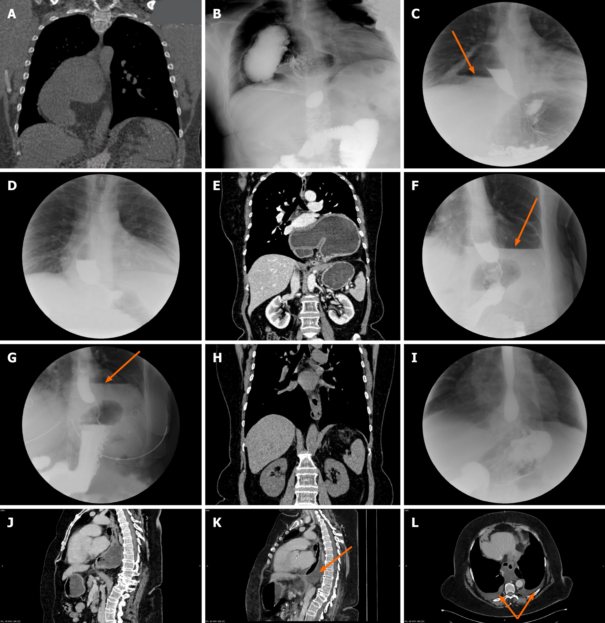
Figure 2 Preoperative and postoperative radiologic images.
A-D: A patient (29-year-old male) in the study group (A: Preoperative computed tomography (CT) scan showing complete displacement of the stomach into the mediastinum and its rotation; B: Preoperative contrast X-ray of the same patient; C: Postoperative contrast X-ray of the patient on the fourth postoperative day after the open procedure. The orange arrow shows the air-fluid level in the mediastinum; D: Postoperative contrast X-ray image of the patient on the 60th postoperative day after the open procedure. There was no air-fluid level left in the mediastinum); E-H: A patient (64-year-old female) in the study group (E: Preoperative CT scan showing complete displacement of the stomach into the mediastinum and its rotation; F: Postoperative contrast X-ray of the patient on the fourth postoperative day after the laparoscopic procedure. The orange arrow shows the air-fluid level in the mediastinum; G: Postoperative contrast X-ray image of the patient on the 25th day after the laparoscopic procedure. The orange arrow shows the air-fluid level in the mediastinum; H: Postoperative CT scan of the patient at 12 months after the laparoscopic procedure. There was no pathologic content left in the mediastinum); I-L: A patient (68-year-old female) in the control group (I: Postoperative X-ray of the patient on the fifth day after laparoscopic hernia repair with removal of the hernia sac; J: Preoperative CT scan showing complete displacement of the stomach into the mediastinum; K: Postoperative CT scan of the patient on the fifth day after laparoscopic hernia repair with removal of the hernia sac. The orange arrow shows the effusion in the mediastinum; L: Postoperative CT scan of the patient on the fifth day after laparoscopic hernia repair with removal of the hernia sac. The orange arrows show the effusion in the pleural cavities). There was no air-fluid level in the mediastinum.
- Citation: Hakobyan VM, Petrosyan AA, Yeghiazaryan HH, Aleksanyan AY, Safaryan HH, Shmavonyan HH, Papazyan KT, Ayvazyan KH, Davtyan LG, Khachatryan AA, Sargsyan GS, Stepanyan SA. Removal of the sac during surgery for the repair of “giant” paraesophageal hernias. World J Gastrointest Surg 2025; 17(7): 106365
- URL: https://www.wjgnet.com/1948-9366/full/v17/i7/106365.htm
- DOI: https://dx.doi.org/10.4240/wjgs.v17.i7.106365


