Copyright
©The Author(s) 2025.
World J Gastrointest Surg. Jul 27, 2025; 17(7): 105503
Published online Jul 27, 2025. doi: 10.4240/wjgs.v17.i7.105503
Published online Jul 27, 2025. doi: 10.4240/wjgs.v17.i7.105503
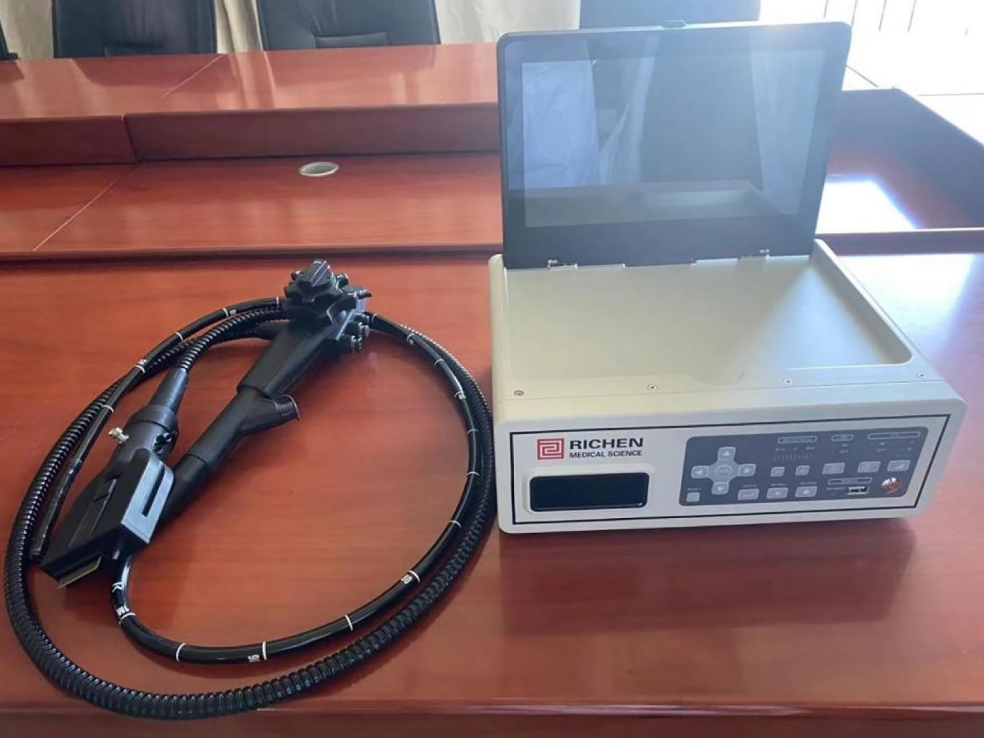
Figure 1 Portable disposable large-channel endoscope system (model HG-GS100).
In this figure, the main components of the portable disposable large-channel endoscope system are shown. The endoscopic host is responsible for powering and controlling the endoscope. The disposable large-channel gastroscope has a distinct structure. The outer diameter of the gastroscope is 9.8 mm, and the channel inner diameter is 3.4 mm with a length of 1450 mm. The tip of the gastroscope contains important components such as the light-emitting diode for illumination, the camera for image capture, and the working channel through which various endoscopic tools can be inserted. The bending section of the endoscope allows for flexible maneuverability during the examination, with an up-bending angle of 180° and down, left, and right bending angles of 160°. These components work in concert to enable effective endoscopic procedures.
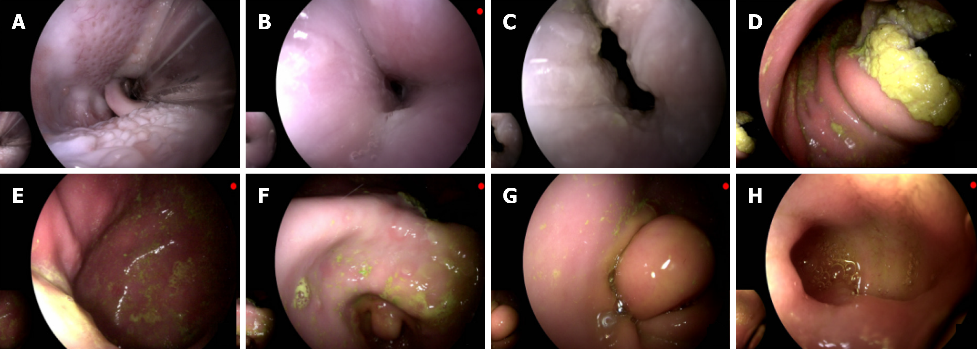
Figure 2 Endoscopic image obtained by the portable disposable large-channel endoscope system.
A: Oropharynx. This area is the initial part of the digestive tract examined during gastroscopy. Clear visualization of the oropharynx is crucial as it can help detect any abnormalities such as inflammation, tumors, or structural defects; B: Esophagus. The esophagus is a key passage for food and liquid transport. Endoscopic examination of the esophagus can identify conditions like esophagitis, esophageal varices, or early-stage cancers. The clear and sharp image of the esophagus obtained by the portable disposable large-channel endoscope system allows for accurate diagnosis; C: Esophagogastric junction. This is the transition area between the esophagus and the stomach. It is an important site for detecting gastroesophageal reflux disease, Barrett's esophagus, and some types of cancer. The image integrity and quality at this junction are essential for proper diagnosis; D: Gastric fundus. The gastric fundus is the upper part of the stomach. It stores food temporarily and plays a role in the initial digestion process. Observing the gastric fundus can help diagnose diseases such as gastric polyps or gastric ulcers; E: Gastric body. The gastric body is the main part of the stomach where most of the digestion and absorption processes occur. Endoscopic evaluation of the gastric body can detect various diseases, and the high-quality image from the endoscope helps in accurate assessment; F: Gastric angle. The gastric angle is a critical anatomical structure for endoscopic examination. It can be a challenging area to visualize clearly, but the portable disposable large-channel endoscope system provides clear images, facilitating the detection of any lesions or abnormalities; G: Gastric antrum. The gastric antrum is involved in the mixing and propulsion of food in the stomach. Abnormalities in this area, such as gastritis or tumors, can be detected through endoscopic images; H: Duodenum. The duodenum is the first part of the small intestine. Endoscopic examination of the duodenum can help diagnose conditions like duodenal ulcers, duodenitis, and some types of tumors. The high-quality images obtained by the endoscope system contribute to accurate diagnosis and treatment planning.
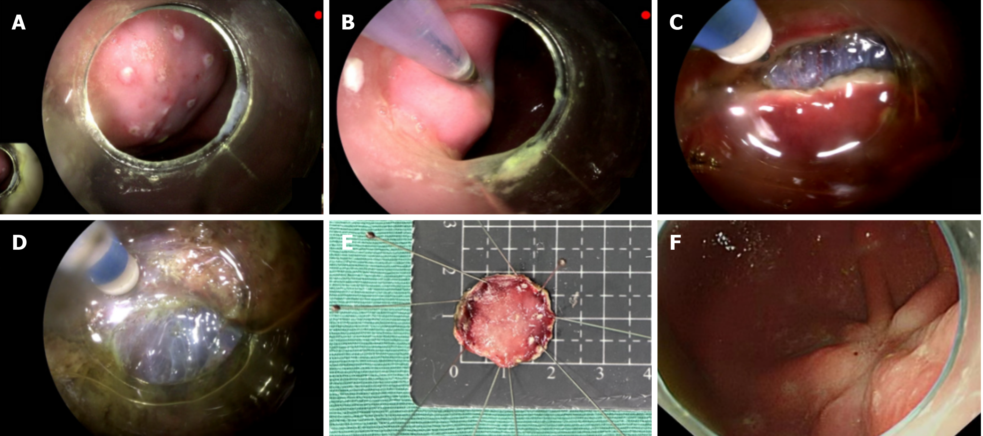
Figure 3 Endoscopic submucosal dissection was performed with the portable disposable large-channel gastroscope.
A: Marking: Marking is the first step in endoscopic submucosal dissection (ESD). It is used to demarcate the boundary of the lesion clearly. This is crucial as it determines the resection margin and ensures complete removal of the lesion. The accurate marking ability of the portable disposable large-channel endoscope system is essential for successful ESD; B: Lifting: Lifting the lesion through submucosal injection is a key step. It separates the lesion from the deeper layers of the digestive tract, making it easier to resect and reducing the risk of perforation. The endoscope's ability to assist in precise lifting is important for the safety and effectiveness of the procedure; C: Circumferential resection: This step involves cutting around the marked lesion. A clear view provided by the endoscope is necessary to ensure a complete and accurate circumferential resection, which is vital for the en-bloc resection of the lesion; D: Dissection: During dissection, the lesion is carefully separated from the underlying tissues. The high-quality image and maneuverability of the portable disposable large-channel endoscope system enable the endoscopist to perform this delicate operation with precision, minimizing the risk of damage to surrounding tissues; E: Specimen: The removed specimen is an important outcome of ESD. The size and integrity of the specimen are used to evaluate the success of the resection and for pathological examination; F: Healing wound: After the ESD procedure, observing the healing wound is crucial. The endoscope can be used to monitor the wound for any signs of bleeding, infection, or improper healing. The clear image quality of the portable disposable large-channel endoscope system helps in accurate wound assessment.
- Citation: Zhao CY, Ning B, Feng XX, Li HK, Zhang WG, Dong H, Chai NL, Linghu EQ. Comparison of a portable disposable large-channel gastroscope and a conventional reusable gastroscope in gastric endoscopic submucosal dissection. World J Gastrointest Surg 2025; 17(7): 105503
- URL: https://www.wjgnet.com/1948-9366/full/v17/i7/105503.htm
- DOI: https://dx.doi.org/10.4240/wjgs.v17.i7.105503


