Copyright
©The Author(s) 2025.
World J Gastrointest Surg. Jul 27, 2025; 17(7): 105308
Published online Jul 27, 2025. doi: 10.4240/wjgs.v17.i7.105308
Published online Jul 27, 2025. doi: 10.4240/wjgs.v17.i7.105308
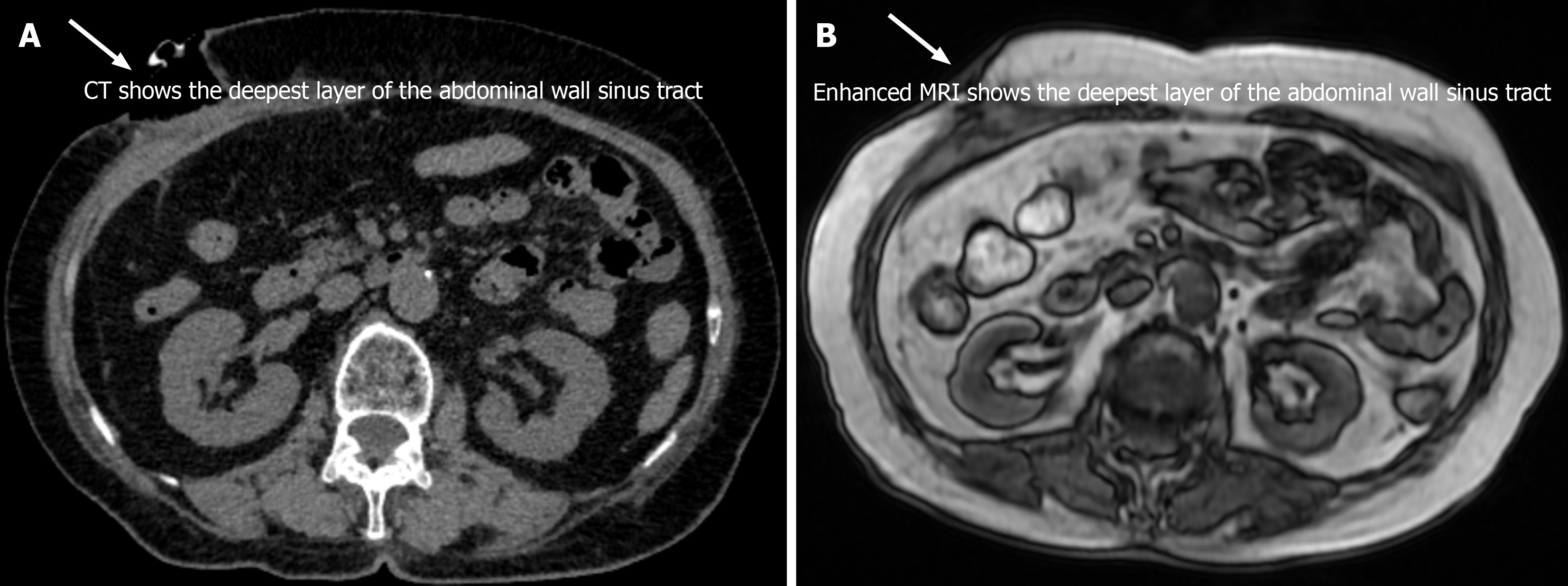
Figure 1 Imaging studies of the abdomen, showing the subcutaneous sinus tract but no definitive connection to the abdominal cavity.
A: Axial computed tomography image showing the deepest layer of the abdominal wall sinus tract. White arrow: Points to the thickened and disordered soft tissue structure in the anterior abdominal wall, indicating the presence of a sinus tract. The image shows that the sinus tract does not extend into the abdominal cavity; B: Coronal magnetic resonance imaging with contrast, emphasizing the deepest layer of the abdominal wall sinus tract on T2-weighted images. White arrow: Points to the deepest layer of the abdominal wall sinus tract in the coronal section. The sinus tract is clearly delineated, confirming its location within the soft tissue layer and its lack of involvement with the internal abdominal structures. This coronal view with contrast further illustrates the thickened and heterogeneous enhancement of the tissue in the upper anterior abdominal wall, confirming the absence of any connection to the abdominal cavity or signs of complications. CT: Computed tomography; MRI: Magnetic resonance imaging.
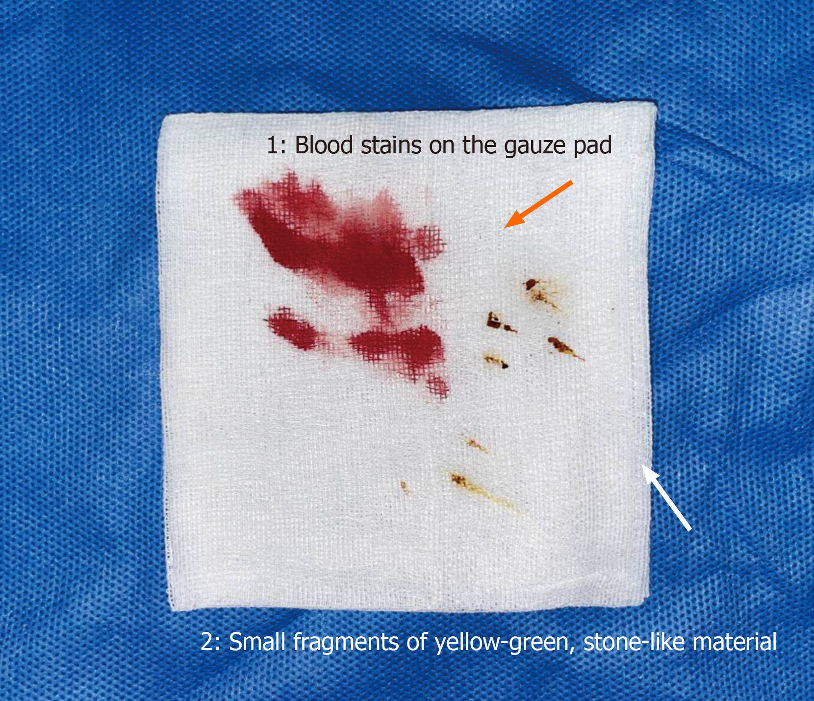
Figure 2 Intraoperative image showing the yellow-green, stone-like material at the base of the sinus tract.
A white gauze pad with blood stains and small fragments of yellow-green material. Orange arrow (1): Blood stains on the gauze pad. Purple arrow (2): Small fragments of yellow-green, stone-like material.
- Citation: Yang L, Wang T, Li XL, Wang YL. Cholecystitis with gallbladder rupture leading to free gallstone migration causing chronic abdominal wall sinus formation: A case report. World J Gastrointest Surg 2025; 17(7): 105308
- URL: https://www.wjgnet.com/1948-9366/full/v17/i7/105308.htm
- DOI: https://dx.doi.org/10.4240/wjgs.v17.i7.105308


