©The Author(s) 2025.
World J Gastrointest Surg. Jun 27, 2025; 17(6): 106009
Published online Jun 27, 2025. doi: 10.4240/wjgs.v17.i6.106009
Published online Jun 27, 2025. doi: 10.4240/wjgs.v17.i6.106009
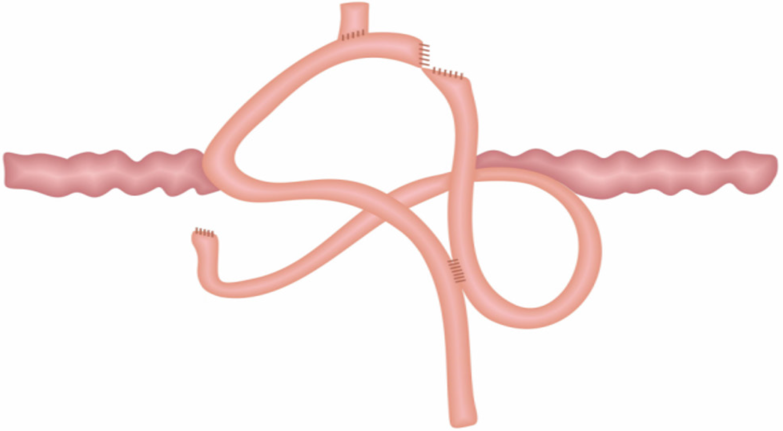
Figure 1 Modified Roux-en-Y digestive tract reconstruction schematic diagram.
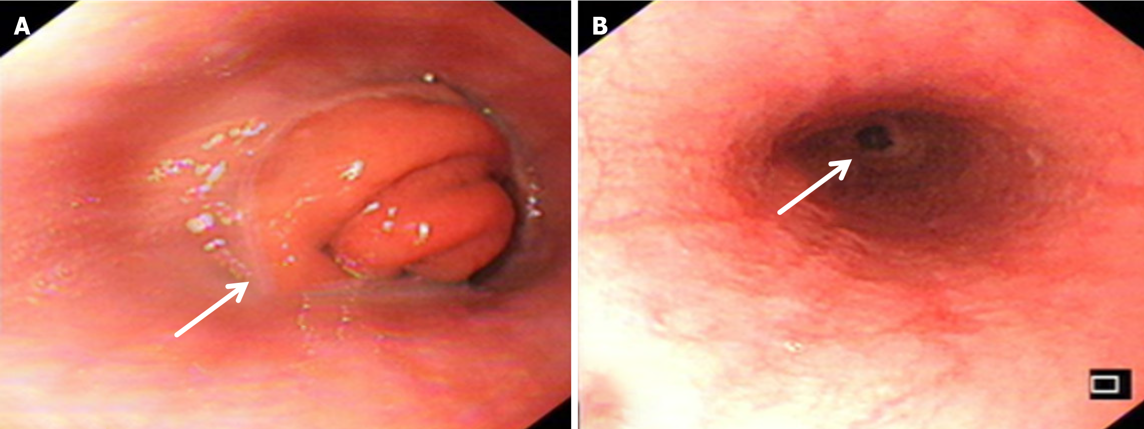
Figure 2 Endoscopic images revealing anastomotic obstruction and stenosis in the Roux-en-Y reconstruction group 1 month after the operation.
A: Digestive endoscopy image showing anastomotic obstruction in the Roux-en-Y reconstruction group at 1 month after the operation; B: Digestive endoscopy image showing anastomotic stenosis in the Roux-en-Y reconstruction group at 1 month after the operation. The arrow indicates the anastomotic situation.
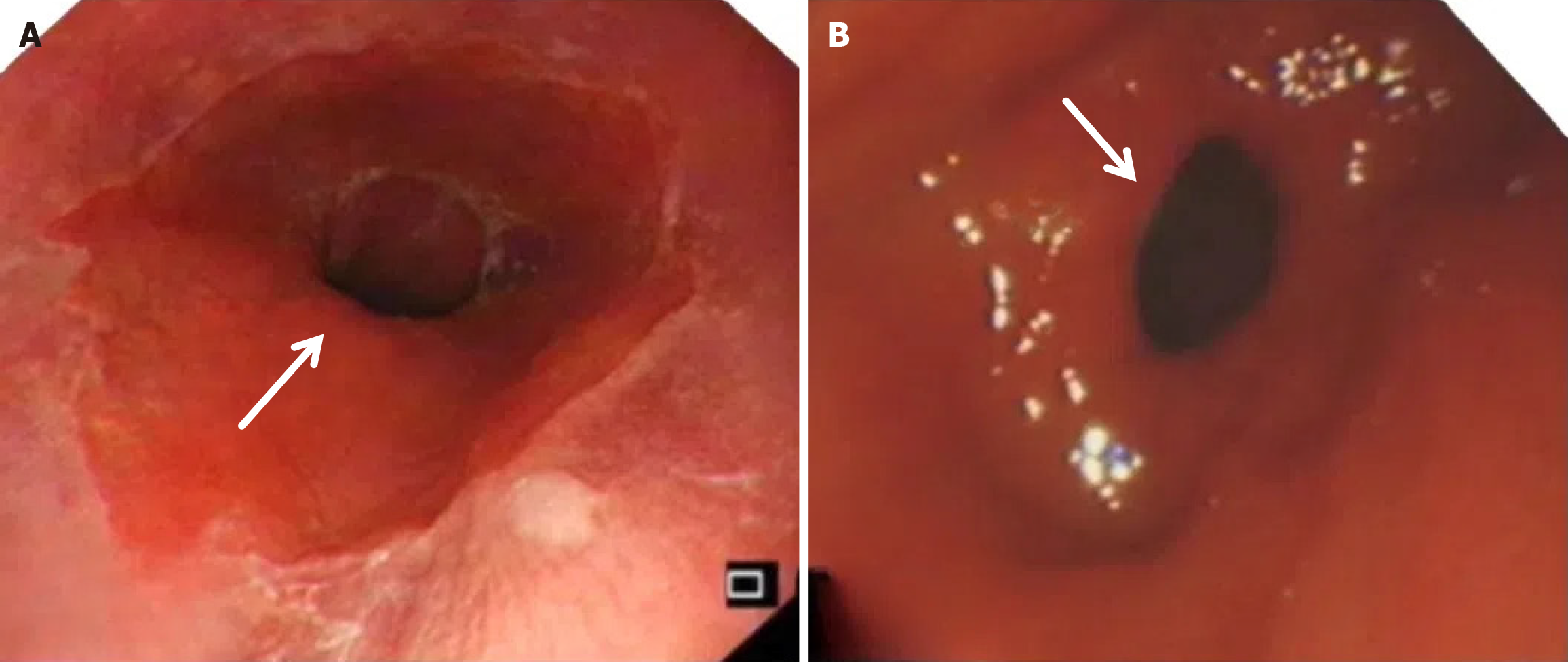
Figure 3 Digestive endoscopy manifestations at 3 months after surgery: Reflux esophagitis in the Roux-en-Y reconstruction group vs the modified group.
A: Digestive endoscopy in the Roux-en-Y reconstruction group revealed reflux esophagitis at 3 months after the operation, and esophageal mucosal congestion and erosion at the anastomotic junction were observed; B: In the modified group, digestive endoscopy revealed that the anastomotic stoma was unobstructed and there was no reflux esophagitis at 3 months after the operation. The arrow indicates the performance of esophageal reflux.
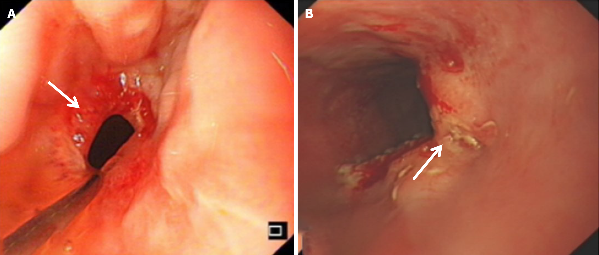
Figure 4 Digestive endoscopic findings of anastomotic complications in the Roux-en-Y reconstruction group at 1 month and 3 months after the operation.
A: In the Roux-en-Y reconstruction group, at 1 month after the operation, digestive endoscopy revealed anastomotic bleeding, and esophageal mucosal bleeding and edema at the anastomotic junction were observed; B: In the Roux-en-Y reconstruction group, digestive endoscopy revealed an anastomotic ulcer, esophageal mucosal erosion, and bleeding at 3 months after the operation. The arrow indicates the anastomotic bleeding and ulcer site.
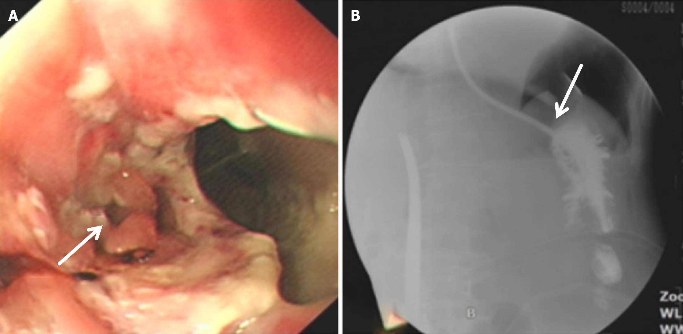
Figure 5 Anastomotic leakage in the Roux-en-Y reconstruction group: Detection by digestive endoscopy and upper gastrointestinal ra
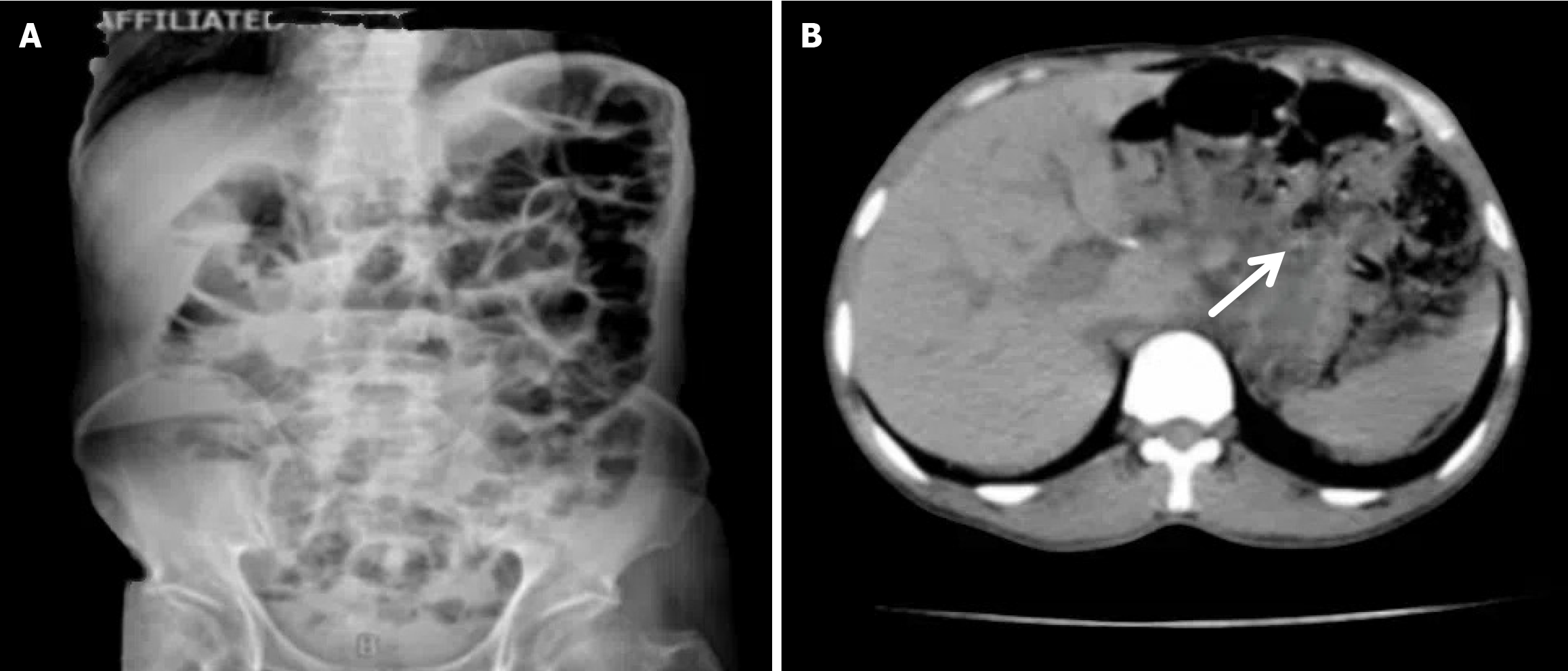
Figure 6 The patient experienced severe abdominal adhesion.
A and B: Abdominal X-ray and abdominal computed tomography images of patients in the Roux-en-Y reconstruction group who were admitted to the hospital due to intestinal obstruction at 3 months after the operation. The patient experienced severe abdominal adhesion, and intraoperative exploration confirmed that it was an abdominal adhesive intestinal obstruction. The arrow indicates the location of intestinal obstruction adhesion zone.
- Citation: Yu J, Li M, Qin XZ, Gong L, Qin L, Lv ZB, Guo W, Huang B, Tian YH. Application of modified Roux-en-Y digestive tract reconstruction in total gastrectomy for patients with gastric cancer. World J Gastrointest Surg 2025; 17(6): 106009
- URL: https://www.wjgnet.com/1948-9366/full/v17/i6/106009.htm
- DOI: https://dx.doi.org/10.4240/wjgs.v17.i6.106009


