Copyright
©The Author(s) 2025.
World J Gastrointest Surg. May 27, 2025; 17(5): 104049
Published online May 27, 2025. doi: 10.4240/wjgs.v17.i5.104049
Published online May 27, 2025. doi: 10.4240/wjgs.v17.i5.104049
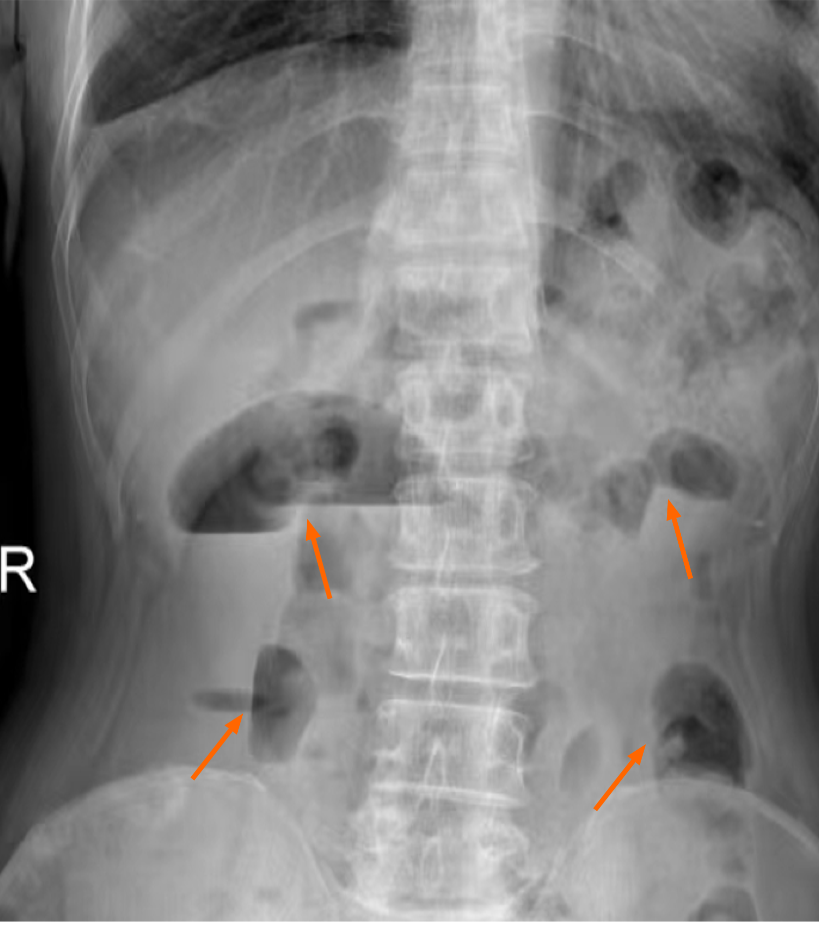
Figure 1 Abdominal upright plain film.
Gas accumulation in the abdominal intestine, and scattered fluid levels are seen, indicating intestinal obstruction.
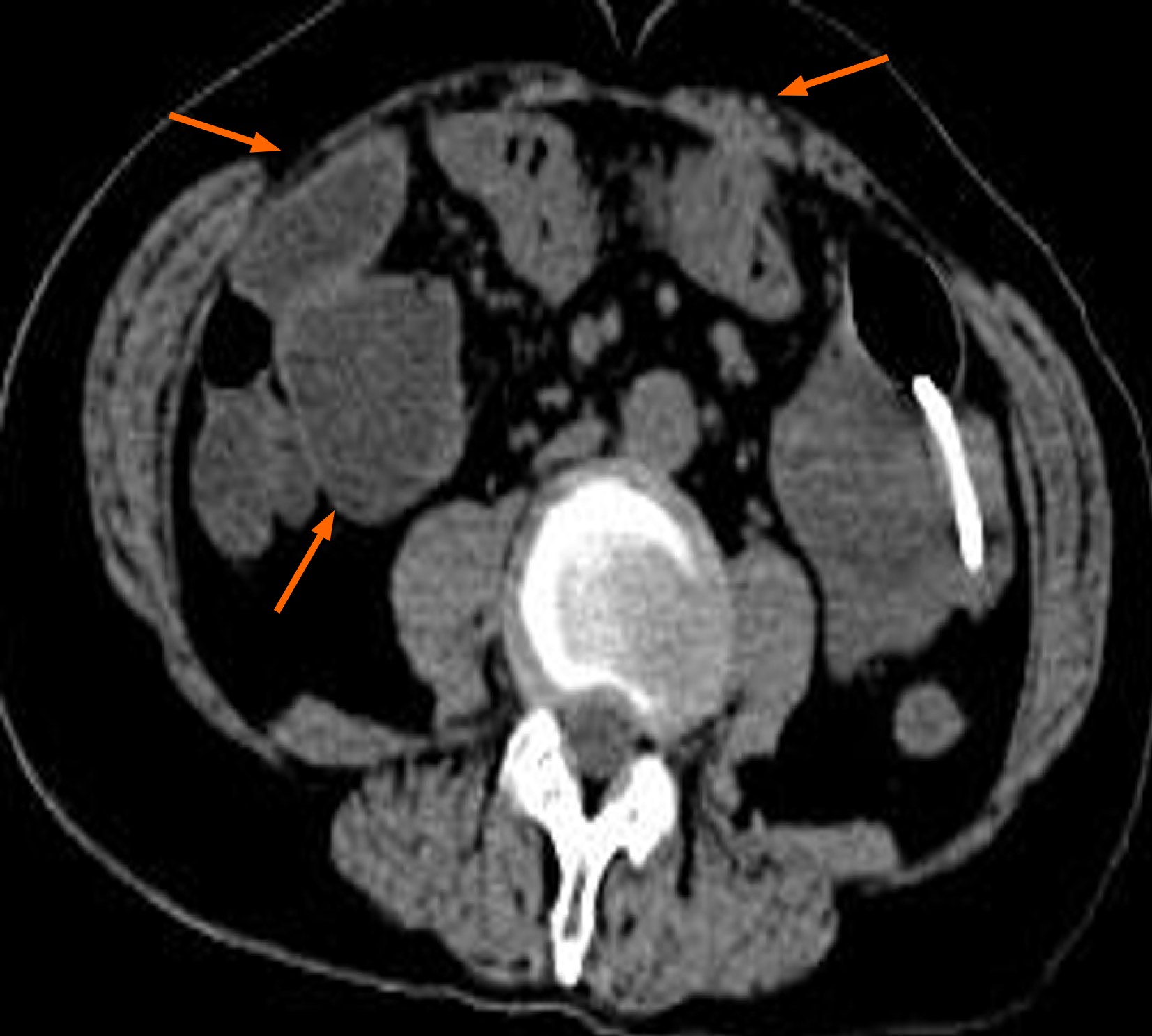
Figure 2 Representative whole-abdominal computed tomography images.
Dilated intestinal collaterals are seen and adhesions of the small intestine to the abdominal wall are seen.
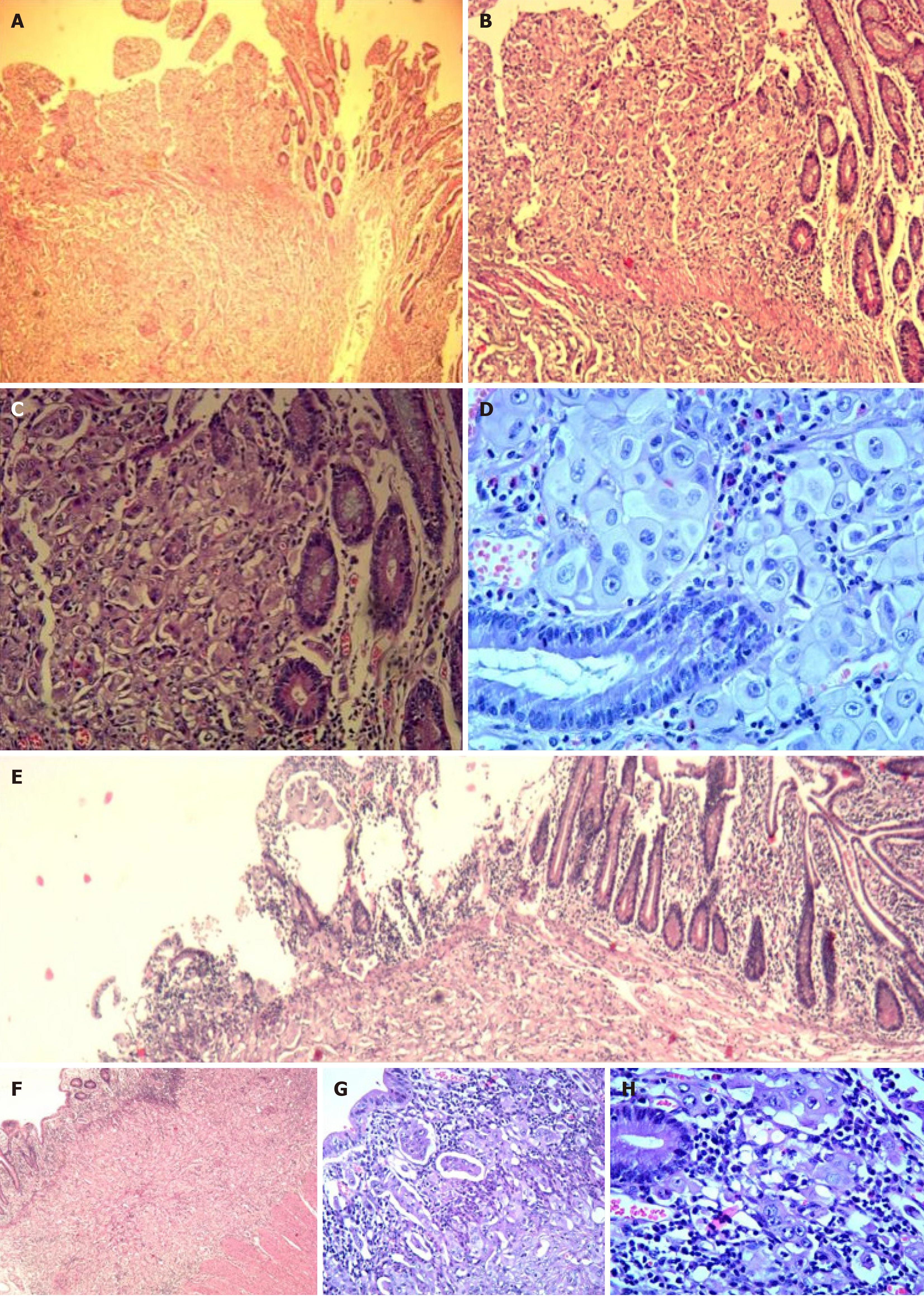
Figure 3 Small intestinal metastasis.
A-D: First small intestine resection specimen. A: Hematoxylin-eosin × 40; B: Hematoxylin-eosin × 100; C: Hematoxylin-eosin × 200; D: Hematoxylin-eosin × 400; E-H: Second small intestine resection specimen (E: Hematoxylin-eosin × 40; F: Hematoxylin-eosin × 100; G: Hematoxylin-eosin × 200; H: Hematoxylin-eosin × 400).
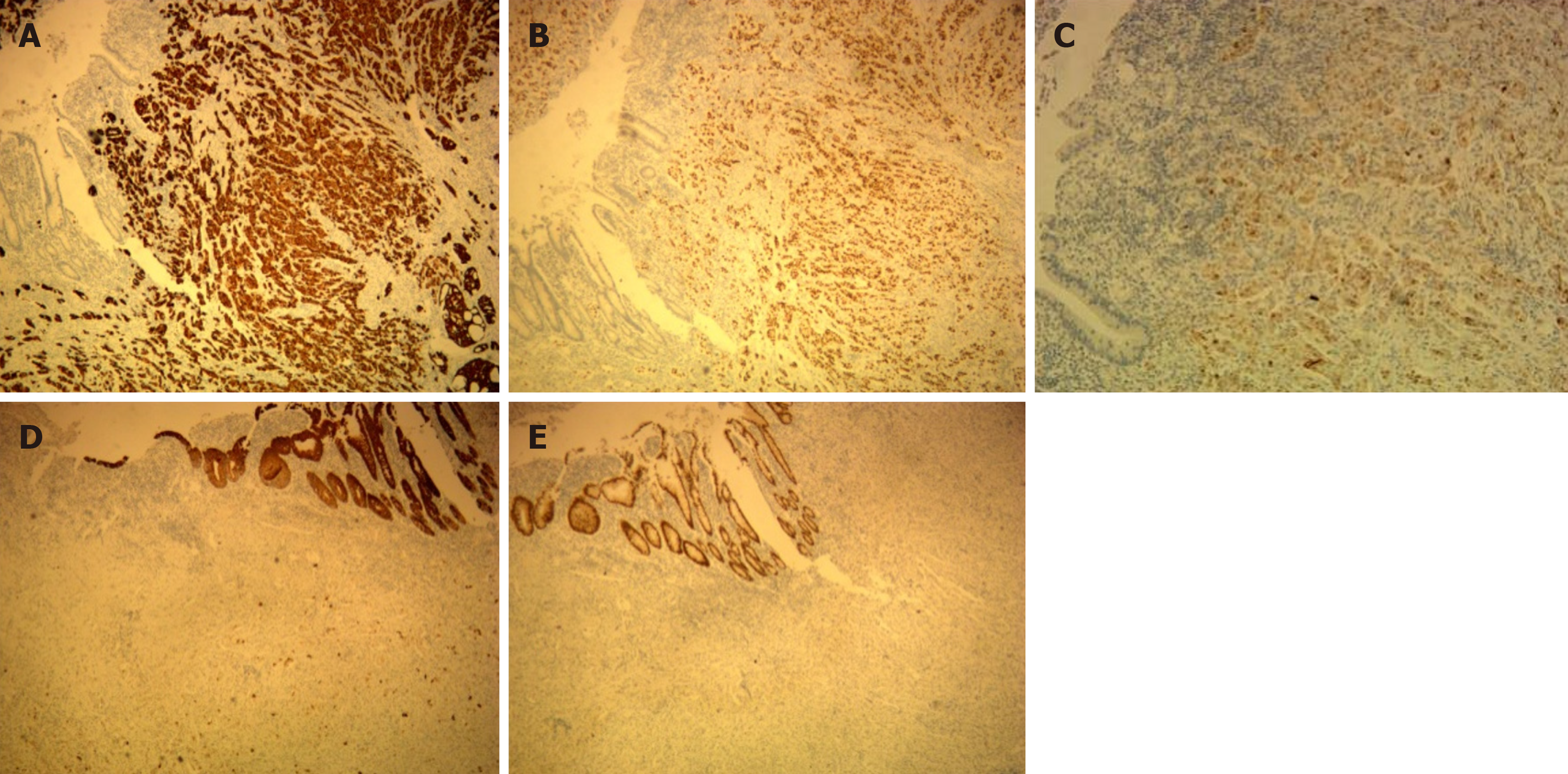
Figure 4 Immunohistochemical tests (first small intestine resection specimen).
A: Cytokeratin (CK) 7 (+++); B: Thyroid transcription factor-1 (++); C: Napsin A (+); D: CK20 (-); E: CDX2 (-).
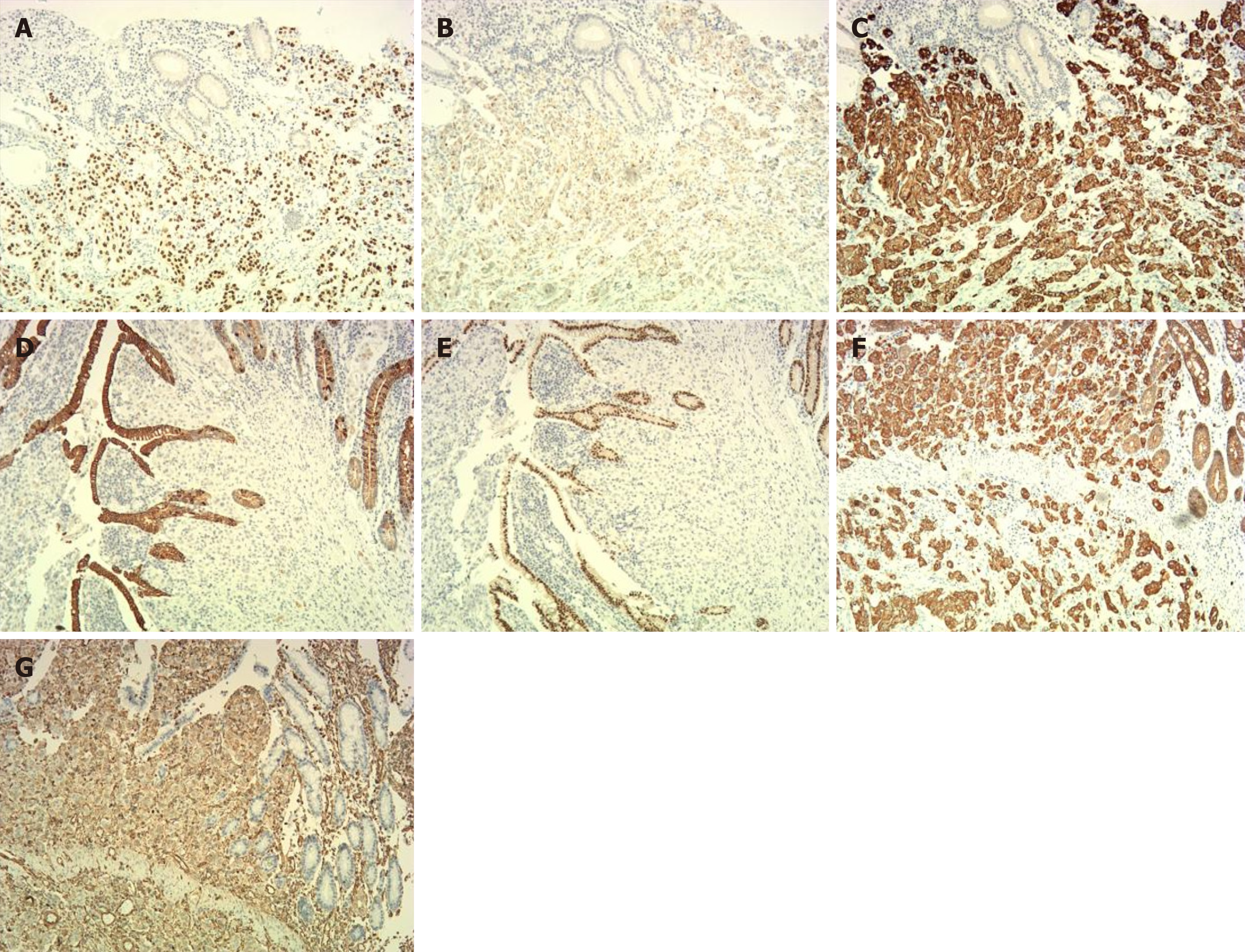
Figure 5 Immunohistochemical tests (second small intestine resection specimen).
A: Thyroid transcription factor-1 (+); B: Napsin-A (partially +); C: Cytokeratin (CK) 7 (+); D: CK20 (partially +); E: CDX2 (-); F: CK-pan (+); G: Vimentin (partially +).
- Citation: Shi RX, Guo ZP, Li X, Wang H, Wang B, Du MY, Wang JJ, Dong ZY. Small intestine metastasis from lung adenocarcinoma: A case report and review of literature. World J Gastrointest Surg 2025; 17(5): 104049
- URL: https://www.wjgnet.com/1948-9366/full/v17/i5/104049.htm
- DOI: https://dx.doi.org/10.4240/wjgs.v17.i5.104049


