©The Author(s) 2025.
World J Gastrointest Surg. Apr 27, 2025; 17(4): 101578
Published online Apr 27, 2025. doi: 10.4240/wjgs.v17.i4.101578
Published online Apr 27, 2025. doi: 10.4240/wjgs.v17.i4.101578
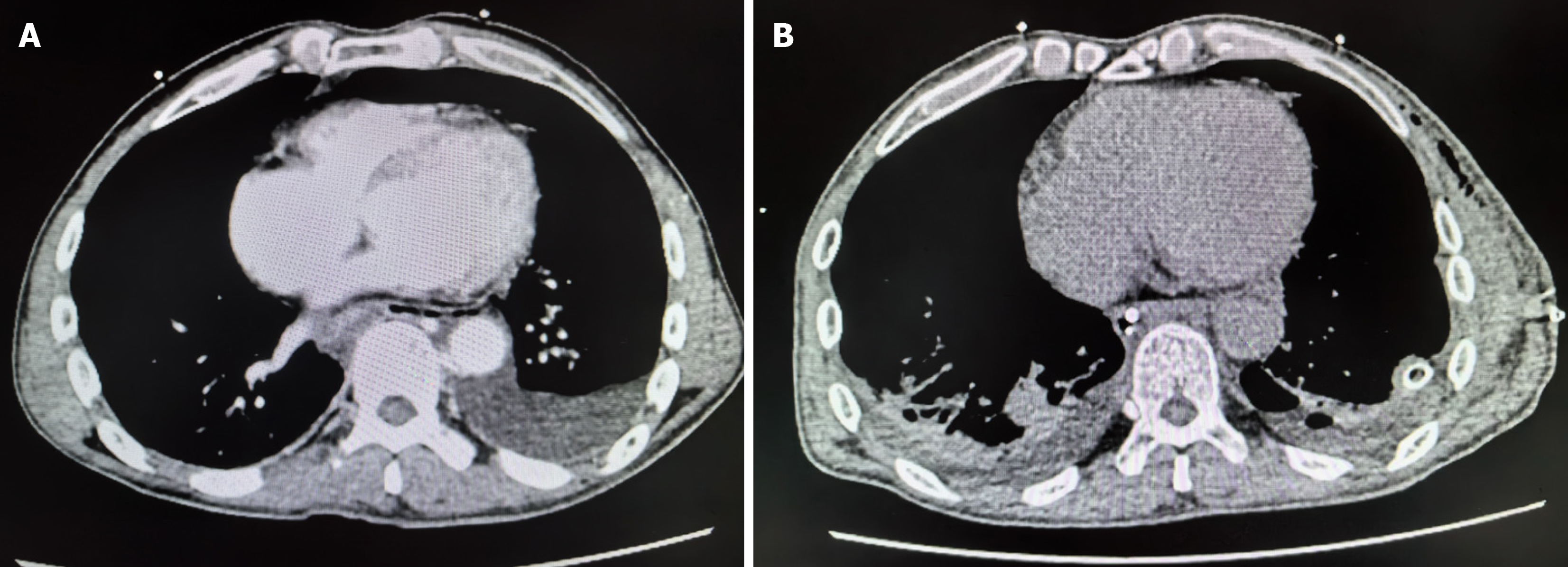
Figure 1 Computed tomography scans.
A: Initial chest and abdominal computed tomography (CT) scans of the patient. CT results indicate a small left-sided pneumothorax caused by severe coughing; approximately 30% compression of the lung tissue is observed; B: Postoperative CT scan. The follow-up CT scan shows a reduction in the left-sided pneumothorax and significant absorption of the mediastinal emphysema, suggesting marked improvement in the thoracic condition.
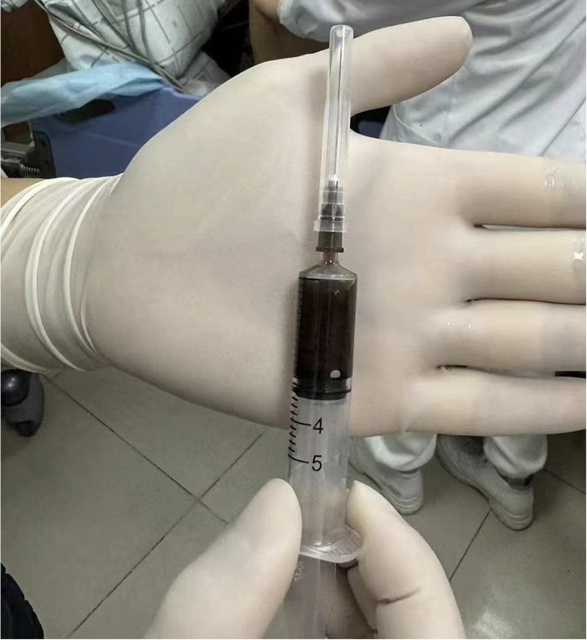
Figure 2 Ultrasound-guided thoracentesis.
Diagnostic thoracentesis was performed under ultrasound guidance, yielding dark brown turbid fluid containing esophageal debris.
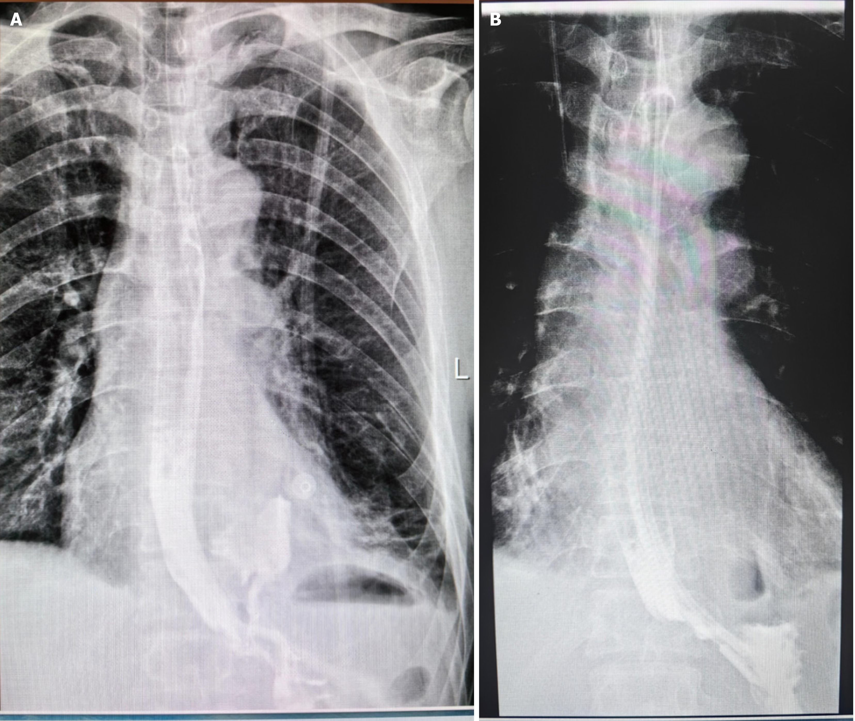
Figure 3 Esophagogram.
A: The esophagogram reveals contrast leakage into the thoracic cavity from the lower esophagus, indicating an esophageal rupture; B: Postoperative esophagogram review. The postoperative esophagogram revealed no contrast leakage, indicating the successful repair of the esophageal perforation.
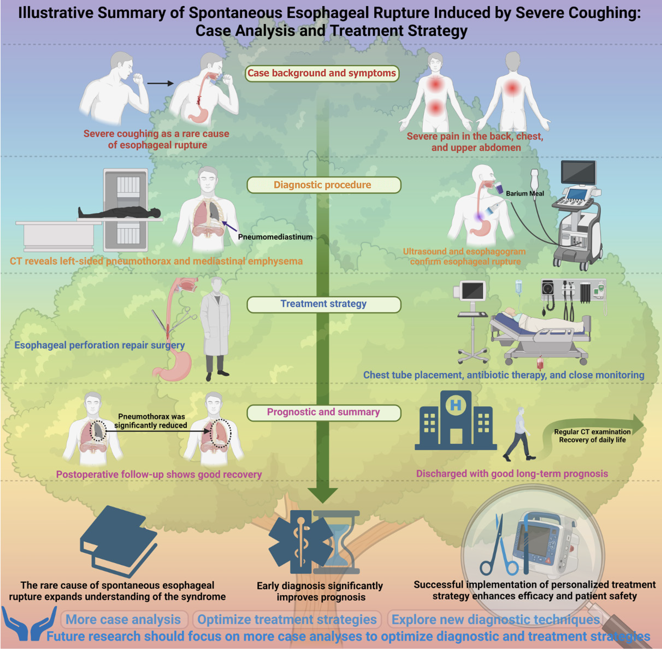
Figure 4 Illustrative summary of spontaneous esophageal rupture induced by severe coughing: Case analysis and treatment strategy.
CT: Computed tomography.
- Citation: Xiong SY, Liu CJ, Li YF, Zhang HL, Chen XW, Wang HM, Chen JC. Multimodal diagnostic and surgical approach to spontaneous esophageal rupture induced by severe coughing: A case report. World J Gastrointest Surg 2025; 17(4): 101578
- URL: https://www.wjgnet.com/1948-9366/full/v17/i4/101578.htm
- DOI: https://dx.doi.org/10.4240/wjgs.v17.i4.101578


