Copyright
©The Author(s) 2025.
World J Gastrointest Surg. Mar 27, 2025; 17(3): 99015
Published online Mar 27, 2025. doi: 10.4240/wjgs.v17.i3.99015
Published online Mar 27, 2025. doi: 10.4240/wjgs.v17.i3.99015
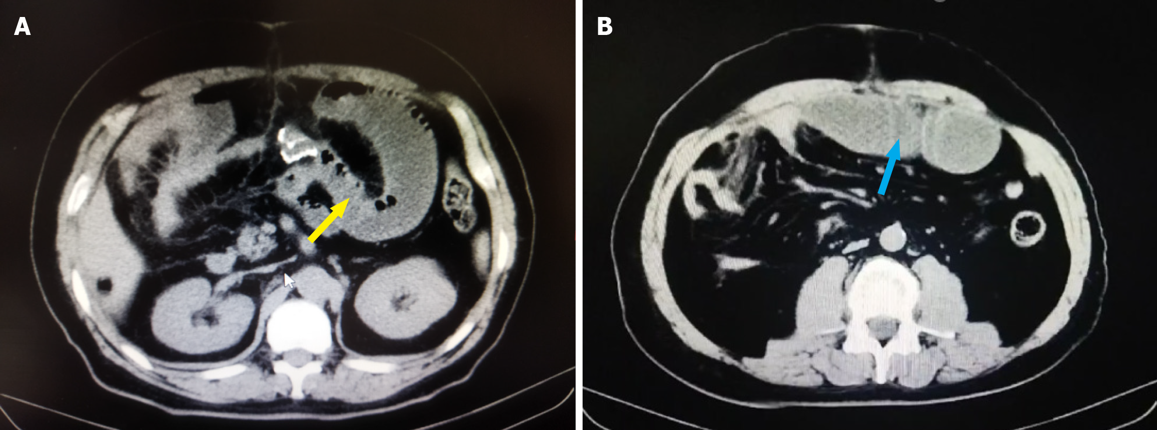
Figure 1 Abdominal computed tomography revealed intestinal obstruction of the patient after abdominal surgery.
A: The yellow arrow indicated the patient's intestinal obstruction at the right renal hilus level; B: The blue arrow indicated the patient’s intestinal obstruction at the umbilical level.
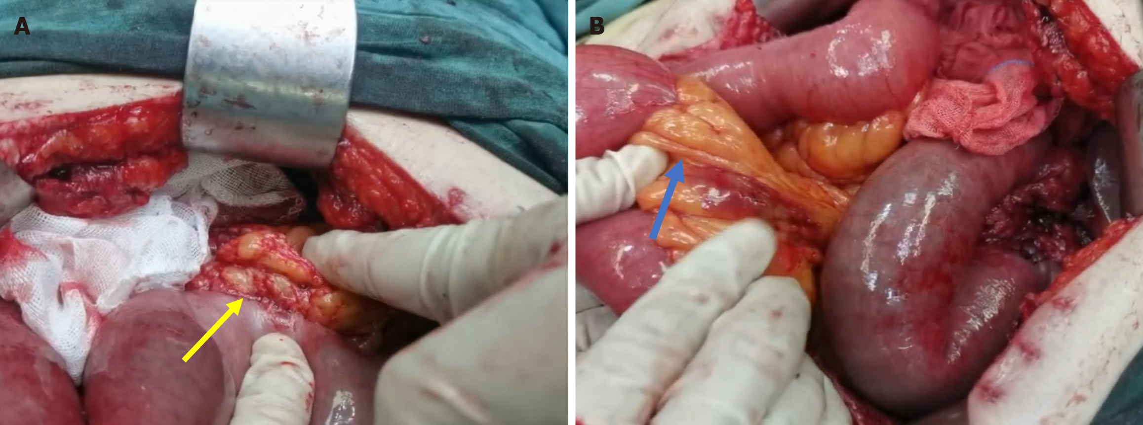
Figure 2 Intraoperative observations may reveal the presence of abdominal adhesions and mesenteric nodules.
A: The yellow arrow indicated a tight adhesion between the mesentery and the small intestine; B: Blue arrow indicated multiple hard nodules in the mesentery.
Figure 3 Pathological findings of surgical specimens.
A and B: Representative hematoxylin and eosin images of mesenteric nodules. Hematoxylin and eosin images showed heterotopic ossification and new bone formation. Black color scale bars: 200μm. The immunohistochemical staining results were as follows: CD68 (+), smooth muscle actin (+), CD34 (+), β-catenin (cytoplasmic+), Ki-67 (+20%), and deamin (-).
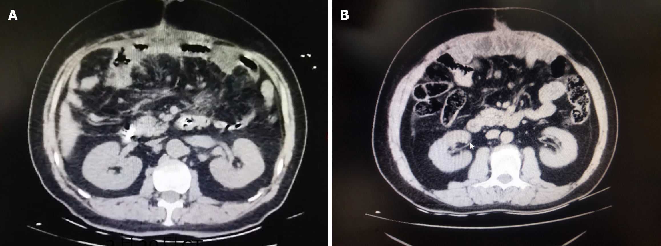
Figure 4 Abdominal computed tomography showed a significant alleviation of intestinal obstruction following methylprednisolone treatment.
A: After 1 week of methylprednisolone treatment, there was a marked reduction in the intestinal obstruction; B: 8 months after methylprednisolone treatment.
- Citation: Chen JT, Li YP, Guo SQ, Huang JS, Wang YG. Nonsurgical treatment of postoperative intestinal obstruction caused by heterotopic ossification of the mesentery: A case report. World J Gastrointest Surg 2025; 17(3): 99015
- URL: https://www.wjgnet.com/1948-9366/full/v17/i3/99015.htm
- DOI: https://dx.doi.org/10.4240/wjgs.v17.i3.99015


